Humerus
Long bone of the arm. It articulates with the scapula at the shoulder and with the radius and ulna at the elbow, and four major nerves lie against it at predictable fracture sites.
Core anatomy
- Location
Arm, from the glenohumeral joint to the elbow.
- Classification
Long bone.
- Key features & landmarks
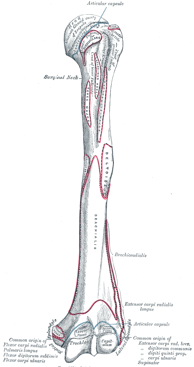
- Head: articulates with the glenoid cavity of the scapula
- Anatomical neck: groove just beyond the head; attachment of the joint capsule
- Surgical neck: narrowing distal to the tubercles; a common fracture site (axillary nerve, posterior circumflex humeral artery)
- Greater tubercle (supraspinatus, infraspinatus, teres minor) and lesser tubercle (subscapularis)
- Intertubercular (bicipital) groove: long head of biceps tendon
- Deltoid tuberosity: insertion of deltoid
- Radial (spiral) groove: posterior shaft; radial nerve and profunda brachii artery
- Medial and lateral epicondyles: common flexor and common extensor origins; the ulnar nerve passes behind the medial epicondyle
- Capitulum (articulates with the radial head) and trochlea (articulates with the ulna)
- Olecranon fossa (posterior), coronoid fossa and radial fossa (anterior)
- Medial and lateral supracondylar ridges
- Relations
Nerves in direct contact with the humerus: axillary nerve (surgical neck), radial nerve (radial groove/shaft), median nerve and brachial artery (anterior to the supracondylar region), ulnar nerve (posterior to the medial epicondyle).
Muscle attachments
- Deltoid — insertion
- Infraspinatus — insertion
- Latissimus dorsi — insertion
- Pectoralis major — insertion
- Pronator teres — origin
- Subscapularis — insertion
- Supraspinatus — insertion
- Teres minor — insertion
- Triceps brachii — origin
Joints
Forms a boundary of
Mentioned in
Generated automatically from links in other entries.

Clinical correlations
Proximal humerus (surgical neck) fracture PANCE
- Mechanism / lesion
Fall on an outstretched hand or onto the shoulder, especially in older women with osteoporosis.
- Classic presentation
Pain, swelling and extensive bruising of the arm and chest wall. Check axillary nerve function (deltoid, regimental badge sensation).
Humeral shaft fracture PANCE
- Mechanism / lesion
Direct blow, fall, or twisting (spiral) injury.
- Classic presentation
Deformity of the arm. Radial nerve palsy (wrist drop) is the classic associated injury, especially with distal-third spiral fractures.
Supracondylar fracture (pediatric) PANCE
- Mechanism / lesion
Most common elbow fracture in children, usually a fall on an outstretched hand with the elbow hyperextended (extension type, distal fragment displaced posteriorly).
- Classic presentation
Elbow pain, swelling and deformity. Assess the brachial artery (radial pulse, perfusion) and the median nerve/AIN, radial and ulnar nerves. Watch for forearm compartment syndrome; untreated ischemia leads to Volkmann ischemic contracture.
Medial epicondyle fracture
- Mechanism / lesion
Valgus stress or avulsion by the flexor–pronator mass, often in adolescents (e.g. with elbow dislocation).
- Classic presentation
Medial elbow pain and swelling; the ulnar nerve is at risk.
High-yield PANCE pearls
- Fracture sites → nerve at risk: surgical neck → axillary, midshaft → radial, supracondylar → median (AIN) + brachial artery, medial epicondyle → ulnar.
- Supracondylar fracture in a child: check pulses, watch for compartment syndrome (pain with passive stretch) → Volkmann contracture.
- Posterior fat pad sign on a lateral elbow film = effusion; in a child with trauma, suspect an occult supracondylar fracture.
- Proximal humerus fracture in an older adult is a fragility fracture; consider osteoporosis evaluation.
Imaging anatomy
- X-ray
Shoulder: AP, scapular Y and axillary views for proximal fractures. Shaft: AP and lateral humerus including both joints. Elbow (pediatric): on a true lateral view, the anterior humeral line should pass through the middle third of the capitellum. In extension-type supracondylar fractures it passes anterior to it. A visible posterior fat pad or elevated anterior fat pad ("sail sign") indicates an effusion. Know the ossification center sequence (CRITOE) so normal centers aren't mistaken for fractures.
- CT
Used for complex proximal humerus or intra-articular distal humerus fractures (surgical planning).
- MRI
Occult fractures, bone bruises, osteonecrosis of the humeral head, and tumors.
- Key views & landmarks
- Shoulder trauma series (AP, scapular Y, axillary)
- Humerus AP and lateral
- Elbow AP and true lateral at 90° (anterior humeral line, fat pads)
Physical exam
- Surface landmarks
The greater tubercle is the most lateral bony point of the shoulder, felt below the acromion. The medial and lateral epicondyles are subcutaneous at the elbow. With the elbow extended, the two epicondyles and the tip of the olecranon lie in a straight line; with the elbow flexed to 90° they form an isosceles triangle. Elbow dislocation disrupts this relationship; a supracondylar fracture typically preserves it.
- Palpation
Palpate the greater tubercle, bicipital groove, shaft (through the muscles) and both epicondyles. Palpate the ulnar nerve behind the medial epicondyle.
- Strength / function testing
In humeral injuries test the axillary nerve (deltoid, lateral shoulder sensation), radial nerve (wrist and finger extension, dorsal first web space), median/AIN ("OK" sign, index pad sensation), ulnar nerve (finger abduction, little finger sensation), and the radial pulse and capillary refill.
Self-quiz
A 6-year-old girl falls from a trampoline onto her outstretched hand. Lateral radiograph of the elbow shows a posterior fat pad, and the anterior humeral line passes anterior to the capitellum. Which structure must be assessed most urgently?
Correct answer: B
These findings indicate an extension-type supracondylar fracture. The brachial artery (and median nerve/AIN) lies directly anterior to the distal humerus and can be injured or kinked. Check the radial pulse and perfusion, and watch for compartment syndrome.
Show answer
B
These findings indicate an extension-type supracondylar fracture. The brachial artery (and median nerve/AIN) lies directly anterior to the distal humerus and can be injured or kinked. Check the radial pulse and perfusion, and watch for compartment syndrome.
Which nerve lies in direct contact with the humerus at the midshaft?
Correct answer: C
The radial nerve lies in the radial (spiral) groove on the posterior midshaft. The axillary nerve is at the surgical neck and the ulnar nerve behind the medial epicondyle.
Show answer
C
The radial nerve lies in the radial (spiral) groove on the posterior midshaft. The axillary nerve is at the surgical neck and the ulnar nerve behind the medial epicondyle.
A 78-year-old woman with osteoporosis falls onto her outstretched arm. Which fracture of the upper limb is she at particular risk of, and which nerve would that fracture endanger?
Correct answer: A
Proximal humerus (surgical neck) fractures are common fragility fractures in older adults. The axillary nerve and posterior circumflex humeral artery wrap around the surgical neck.
Show answer
A
Proximal humerus (surgical neck) fractures are common fragility fractures in older adults. The axillary nerve and posterior circumflex humeral artery wrap around the surgical neck.
With the elbow flexed to 90°, the medial epicondyle, lateral epicondyle and olecranon normally form which configuration?
Correct answer: B
In flexion the three points form an isosceles triangle; in extension they lie in a straight line. A posterior elbow dislocation disrupts this relationship.
Show answer
B
In flexion the three points form an isosceles triangle; in extension they lie in a straight line. A posterior elbow dislocation disrupts this relationship.
A 13-year-old baseball pitcher has a displaced avulsion fracture of the medial epicondyle. Which nerve is most at risk?
Correct answer: D
The ulnar nerve passes behind the medial epicondyle, so medial epicondyle fractures threaten it. Other fracture–nerve pairs: surgical neck → axillary; midshaft → radial; supracondylar → median (AIN) and brachial artery.
Show answer
D
The ulnar nerve passes behind the medial epicondyle, so medial epicondyle fractures threaten it. Other fracture–nerve pairs: surgical neck → axillary; midshaft → radial; supracondylar → median (AIN) and brachial artery.