Cranial meninges and intracranial hemorrhage
Three membranes — dura, arachnoid and pia — that cover the brain and define the epidural, subdural and subarachnoid spaces. Each space has its own classic bleed and CT appearance.
Core anatomy
- Location
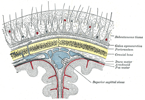
Between the skull and the brain; continuous through the foramen magnum with the spinal meninges and spaces.
- Boundaries
- dura mater
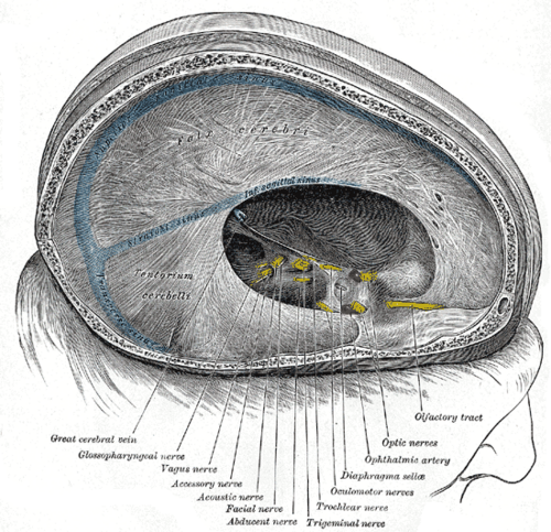
- Tough outer layer with a periosteal layer (fused to the skull) and a meningeal layer; they separate to enclose the dural venous sinuses and fold inward as the falx cerebri (between the hemispheres), tentorium cerebelli (over the cerebellum), falx cerebelli and diaphragma sellae
- arachnoid mater
- Thin avascular layer under the dura; arachnoid granulations project into the sinuses
- pia mater
- Delicate layer adherent to the brain surface, following every gyrus
- Contents
- Epidural space (potential): between skull and dura — contains the middle meningeal artery and meningeal veins
- Subdural space (potential): between dura and arachnoid — crossed by bridging veins from the cortex to the sinuses
- Subarachnoid space: between arachnoid and pia — CSF, the cerebral arteries of the circle of Willis, and enlargements called cisterns
- Communications
The subarachnoid space is continuous with the ventricles via the fourth ventricle apertures and with the spinal subarachnoid space (see ventricular system and CSF).
- Notes
The dura is pain-sensitive: supratentorial dura is supplied by the trigeminal nerve and infratentorial dura by C2–C3 (and CN X) — which is why posterior fossa lesions cause occipital/neck pain. The brain parenchyma itself has no pain fibers.
Mentioned in
Generated automatically from links in other entries.
Swipe sideways to see the whole diagram →



Clinical correlations
Epidural hematoma PANCE
- Mechanism / lesion
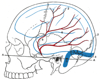
Arterial bleeding, classically the middle meningeal artery torn by a temporal bone (pterion) fracture.
- Classic presentation
Head injury with brief loss of consciousness, a lucid interval, then rapid deterioration (headache, vomiting, ipsilateral blown pupil, contralateral hemiparesis — uncal herniation). CT: biconvex (lens-shaped) hyperdensity that does not cross suture lines. Emergency craniotomy.
Subdural hematoma PANCE
- Mechanism / lesion
Tearing of bridging veins — elderly (brain atrophy), alcohol use disorder, anticoagulation, falls; infants (abusive head trauma).
- Classic presentation
Acute (after major trauma) or chronic (weeks after minor trauma — gradual headache, confusion, fluctuating consciousness, mild hemiparesis). CT: crescent-shaped collection that crosses sutures but not the falx; hyperdense acute, isodense subacute, hypodense chronic. Shaken infants: subdural hematomas + retinal hemorrhages.
Subarachnoid hemorrhage PANCE
- Mechanism / lesion
Rupture of a saccular (berry) aneurysm of the circle of Willis (most nontraumatic cases); also trauma and AVMs. Risks — hypertension, smoking, ADPKD, connective tissue disorders, family history.
- Classic presentation
Sudden "thunderclap" headache ("worst of my life"), vomiting, neck stiffness, photophobia, decreased consciousness. Noncontrast CT (very sensitive within 6 hours) → if negative, lumbar puncture (xanthochromia, RBCs that do not clear) → CTA to find the aneurysm. Complications: rebleeding, vasospasm (days 3–14 — nimodipine), hydrocephalus, hyponatremia. Secure the aneurysm (coiling or clipping).
Bacterial meningitis PANCE
- Mechanism / lesion
Infection of the leptomeninges and subarachnoid space — S. pneumoniae, N. meningitidis, Listeria (age > 50, immunocompromised, neonates), GBS and E. coli in neonates.
- Classic presentation
Fever, headache, neck stiffness, altered mental status, petechial rash (meningococcus). CSF: high opening pressure, neutrophils, low glucose, high protein. Give dexamethasone and empiric antibiotics (ceftriaxone + vancomycin ± ampicillin) immediately — do not delay for CT or LP.
High-yield PANCE pearls
- Lucid interval + lens-shaped bleed not crossing sutures → epidural (middle meningeal artery).
- Crescent crossing sutures in an elderly/alcoholic/anticoagulated patient → subdural (bridging veins).
- Thunderclap headache → noncontrast CT → if negative, LP for xanthochromia → SAH (berry aneurysm).
- SAH → nimodipine to prevent vasospasm.
- Meningitis: neutrophils + low glucose + high protein → bacterial → antibiotics + dexamethasone without delay.
Imaging anatomy
- CT
Noncontrast CT head — first test for trauma and suspected hemorrhage (epidural = biconvex; subdural = crescent; SAH = blood in the basal cisterns and sulci).
- MRI
MRI for subacute/chronic subdural collections, small SAH (FLAIR), meningeal enhancement in meningitis.
- Key views & landmarks
- CT angiography for aneurysm after SAH
- Lumbar puncture: opening pressure, cells, glucose, protein, Gram stain, xanthochromia
Physical exam
- Surface landmarks
The pterion (junction of frontal, parietal, temporal and sphenoid bones, about 3 cm above the zygomatic arch midpoint) overlies the anterior branch of the middle meningeal artery.
- Strength / function testing
Glasgow Coma Scale, pupils, focal deficits; meningeal signs — neck stiffness, Kernig (pain on extending the knee with the hip flexed) and Brudzinski (neck flexion causes hip flexion) signs, which are specific but insensitive.
Self-quiz
A 19-year-old is hit in the temple by a baseball, is briefly unconscious, then talks normally for 2 hours before becoming drowsy with a dilated right pupil. CT shows a lens-shaped hyperdensity. Which vessel is most likely injured?
Correct answer: B
A lucid interval and a biconvex (lens-shaped) collection after a temporal blow indicate an epidural hematoma from the middle meningeal artery — a neurosurgical emergency.
Show answer
B
A lucid interval and a biconvex (lens-shaped) collection after a temporal blow indicate an epidural hematoma from the middle meningeal artery — a neurosurgical emergency.
A 45-year-old has a sudden severe headache during exercise. Noncontrast CT 10 hours later is normal. What is the next step?
Correct answer: B
A thunderclap headache needs subarachnoid hemorrhage excluded. If CT is negative (especially after 6 hours), perform an LP looking for xanthochromia and persistent RBCs (or CTA per local protocol).
Show answer
B
A thunderclap headache needs subarachnoid hemorrhage excluded. If CT is negative (especially after 6 hours), perform an LP looking for xanthochromia and persistent RBCs (or CTA per local protocol).
An 82-year-old on warfarin has had increasing confusion for 3 weeks after a minor fall. CT shows a crescent-shaped hypodense collection over the left hemisphere that crosses suture lines. What is the diagnosis?
Correct answer: B
A crescent collection crossing sutures is subdural; hypodensity after weeks means it is chronic. Elderly anticoagulated patients are at high risk from bridging vein tears.
Show answer
B
A crescent collection crossing sutures is subdural; hypodensity after weeks means it is chronic. Elderly anticoagulated patients are at high risk from bridging vein tears.
Which drug is given after aneurysmal subarachnoid hemorrhage to reduce the risk of ischemia from vasospasm?
Correct answer: A
Nimodipine is given after SAH to reduce ischemic complications of vasospasm, which peaks at days 3–14.
Show answer
A
Nimodipine is given after SAH to reduce ischemic complications of vasospasm, which peaks at days 3–14.
Which nerve carries pain from the supratentorial dura?
Correct answer: D
The supratentorial dura is supplied by the trigeminal nerve; the infratentorial dura by C2–C3 (and CN X), which is why posterior fossa lesions cause occipital and neck pain. The brain itself has no pain fibers.
Show answer
D
The supratentorial dura is supplied by the trigeminal nerve; the infratentorial dura by C2–C3 (and CN X), which is why posterior fossa lesions cause occipital and neck pain. The brain itself has no pain fibers.