Wrist joint
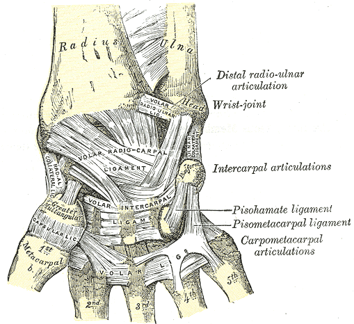
Condyloid joint between the distal radius (and articular disc) and the proximal carpal row. It's a common site of fractures, instability and tenosynovitis.
Core anatomy
- Location
Between the distal forearm and the carpus.
- Classification
Synovial condyloid (ellipsoid), biaxial joint.
- Articulating surfaces
Distal radius and the articular disc of the triangular fibrocartilage complex (TFCC) proximally, with the scaphoid, lunate and triquetrum distally. The ulna does not articulate directly with the carpus; the articular disc separates it.
- Capsule
Fibrous capsule reinforced by the palmar and dorsal radiocarpal ligaments and the ulnar and radial collateral ligaments.
- Ligaments
- Palmar radiocarpal and palmar ulnocarpal ligaments (strong)
- Dorsal radiocarpal ligament
- Radial and ulnar collateral ligaments of the wrist
- Scapholunate and lunotriquetral interosseous ligaments (key to carpal stability)
- TFCC: articular disc, radioulnar ligaments and ulnocarpal ligaments; stabilizes the DRUJ and cushions the ulnar carpus
- Movements
- Flexion and extension (shared with the midcarpal joint)
- Abduction (radial deviation) and adduction (ulnar deviation)
- Circumduction
- Stability
Carpal ligaments, the TFCC and the tendons crossing the wrist.
- Innervation
Anterior interosseous branch of the median nerve, posterior interosseous branch of the radial nerve, and dorsal and deep branches of the ulnar nerve
- Blood supply
Dorsal and palmar carpal arches from the radial artery and ulnar artery
- Relations
Volar: the carpal tunnel (median nerve and flexor tendons) and Guyon's canal (ulnar nerve and artery). Dorsal: the extensor tendons in six compartments; the first compartment (APL, EPB) is the site of de Quervain tenosynovitis. Radial: the anatomical snuffbox with the radial artery.
Swipe sideways to see the whole diagram →

Clinical correlations
De Quervain tenosynovitis PANCE
- Mechanism / lesion
Stenosing tenosynovitis of the first dorsal compartment (abductor pollicis longus and extensor pollicis brevis) from repetitive thumb and wrist motion, e.g. new parents lifting infants.
- Classic presentation
Radial-sided wrist pain and tenderness over the radial styloid, with a positive Finkelstein test.
Ganglion cyst PANCE
- Mechanism / lesion
Mucin-filled cyst arising from a joint capsule or tendon sheath. It is the most common soft-tissue mass of the hand and wrist, most often dorsal over the scapholunate region.
- Classic presentation
Smooth, firm, often transilluminating mass that may change in size. Usually painless or mildly painful.
Lunate and perilunate dislocation
- Mechanism / lesion
High-energy hyperextension (fall from height, motor vehicle collision).
- Classic presentation
Wrist pain and swelling. The volarly displaced lunate may compress the median nerve (acute carpal tunnel syndrome). Easily missed without a true lateral radiograph.
Kienböck disease
- Mechanism / lesion
Avascular necrosis of the lunate, associated with negative ulnar variance (a short ulna).
- Classic presentation
Dorsal wrist pain and stiffness. Progressive sclerosis and collapse of the lunate.
TFCC injury
- Mechanism / lesion
Fall on an outstretched pronated hand, or degenerative (positive ulnar variance).
- Classic presentation
Ulnar-sided wrist pain, clicking, worse with ulnar deviation and rotation.
High-yield PANCE pearls
- De Quervain: APL + EPB (first dorsal compartment); Finkelstein positive; new parents.
- Ganglion cyst: most common hand/wrist mass; dorsal; transilluminates.
- Lunate dislocation → acute median nerve compression; "spilled teacup" on lateral X-ray.
- Proximal row (lateral to medial): scaphoid, lunate, triquetrum, pisiform. Distal row: trapezium, trapezoid, capitate, hamate.
Imaging anatomy
- X-ray
PA, lateral and oblique. PA: Gilula arcs (three smooth arcs along the carpal rows) should be unbroken; scapholunate gap (widening suggests scapholunate dissociation); ulnar variance. Lateral: the radius, lunate and capitate should be colinear. In lunate dislocation, the lunate tips volarly ("spilled teacup") and looks triangular on PA ("piece of pie").
- CT
Occult carpal fractures and fracture characterization.
- MRI
TFCC tears (MR arthrography improves sensitivity), ligament injuries, Kienböck disease, occult scaphoid fracture.
- Ultrasound
Ganglion cysts (anechoic, well defined), de Quervain tenosynovitis (thickened first-compartment tendons and sheath), and guidance for injections.
- Key views & landmarks
- Wrist PA, lateral and oblique
- Scaphoid view (PA ulnar deviation)
- MR arthrogram for TFCC
Physical exam
- Surface landmarks
Radial styloid, Lister's tubercle, ulnar head and styloid, anatomical snuffbox (scaphoid), pisiform (base of the hypothenar eminence), hook of hamate (in the hypothenar eminence, distal and radial to the pisiform), and the scaphoid tubercle (base of the thenar eminence).
- Palpation
Palpate systematically radial → dorsal → ulnar → volar, including the snuffbox, first dorsal compartment, TFCC (ulnar fovea) and pisiform.
- Strength / function testing
Active and passive flexion, extension, and radial and ulnar deviation. Grip strength.
Finkelstein test
- Technique
The patient flexes the thumb into the palm and closes the fingers over it, then the examiner ulnarly deviates the wrist. (This commonly taught form is technically the Eichhoff maneuver; in Finkelstein's original, the examiner grasps the thumb and deviates the wrist.)
- Positive test
Sharp pain over the radial styloid (first dorsal compartment).
- Significance
De Quervain tenosynovitis. The commonly taught form produces false positives in normal wrists.
Ulnar fovea sign
- Technique
Press into the soft spot between the ulnar styloid and FCU tendon, between the pisiform and the ulnar head.
- Positive test
Tenderness reproducing the patient's pain.
- Significance
Suggests a TFCC foveal tear or ulnotriquetral ligament injury. verify
Watson scaphoid shift test
- Technique
The examiner presses on the scaphoid tubercle while moving the wrist from ulnar to radial deviation.
- Positive test
Pain and/or a clunk.
- Significance
Scapholunate instability.
Self-quiz
A 31-year-old woman, 6 weeks postpartum, has pain over the radial side of her wrist when lifting her baby. Pain is reproduced when she makes a fist around her thumb and ulnarly deviates the wrist. Which tendons are involved?
Correct answer: B
De Quervain tenosynovitis involves the first dorsal compartment: APL and EPB. EPL is in the third compartment, where it turns around Lister's tubercle.
Show answer
B
De Quervain tenosynovitis involves the first dorsal compartment: APL and EPB. EPL is in the third compartment, where it turns around Lister's tubercle.
Which bones form the distal articular surface of the radiocarpal joint?
Correct answer: A
The radius and articular disc articulate with the scaphoid, lunate and triquetrum. The pisiform sits on the triquetrum and isn't part of the radiocarpal joint.
Show answer
A
The radius and articular disc articulate with the scaphoid, lunate and triquetrum. The pisiform sits on the triquetrum and isn't part of the radiocarpal joint.
After a fall from a ladder, a patient has wrist pain and numbness of the thumb, index and middle fingers. The lateral radiograph shows the lunate tipped volarly, like a spilled teacup. Which nerve is at risk?
Correct answer: B
A volarly dislocated lunate pushes into the carpal tunnel and compresses the median nerve (acute carpal tunnel syndrome). This is an orthopedic emergency.
Show answer
B
A volarly dislocated lunate pushes into the carpal tunnel and compresses the median nerve (acute carpal tunnel syndrome). This is an orthopedic emergency.
Kienböck disease is avascular necrosis of which bone, and which anatomic variant is associated with it?
Correct answer: B
Kienböck disease is AVN of the lunate, associated with negative ulnar variance. Positive ulnar variance is associated with degenerative TFCC injury. Scaphoid AVN follows fracture.
Show answer
B
Kienböck disease is AVN of the lunate, associated with negative ulnar variance. Positive ulnar variance is associated with degenerative TFCC injury. Scaphoid AVN follows fracture.
A 26-year-old has a smooth, firm, painless mass on the dorsal wrist over the scapholunate region. It transilluminates and varies in size. What is the most likely diagnosis?
Correct answer: A
A ganglion cyst is the most common soft-tissue mass of the hand and wrist, usually dorsal over the scapholunate region. It is mucin-filled, arises from a joint capsule or tendon sheath, often transilluminates and changes in size.
Show answer
A
A ganglion cyst is the most common soft-tissue mass of the hand and wrist, usually dorsal over the scapholunate region. It is mucin-filled, arises from a joint capsule or tendon sheath, often transilluminates and changes in size.
Flashcards
See also
- Scaphoid
- Radius
- Carpal tunnel
- Fall on an outstretched hand (clinical case)
- Numb fingers that wake her at night (clinical case)