Testis and epididymis
Paired male gonads in the scrotum with the epididymis along their back. Torsion is a surgical emergency, and aortic blood supply plus para-aortic lymph drainage reflect the testis's descent from the abdomen.
Core anatomy
- Location
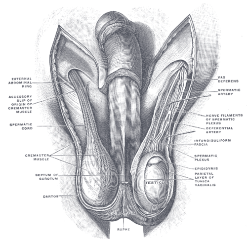
In the scrotum, suspended by the spermatic cord. Each testis is about 4–5 cm long; the left usually hangs slightly lower.
- Parts
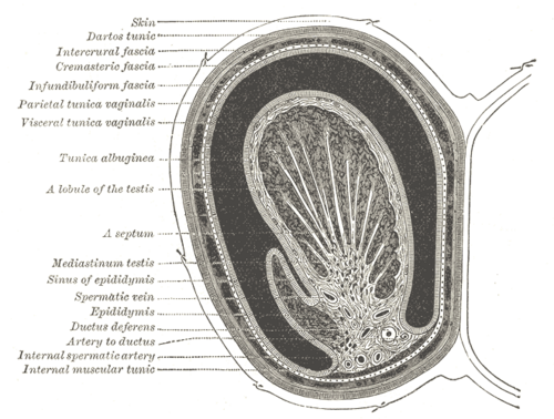
- Tunica vaginalis: closed peritoneal sac (from the processus vaginalis) with parietal and visceral layers covering the front and sides of the testis
- Tunica albuginea: tough fibrous capsule; septa divide the testis into lobules of seminiferous tubules (spermatogenesis; Sertoli cells) with Leydig cells (testosterone) between them
- Seminiferous tubules → straight tubules → rete testis → efferent ductules → epididymis
- Epididymis: head (upper pole), body and tail along the posterolateral testis; stores and matures sperm; the tail continues as the ductus deferens and seminal glands
- Appendix testis and appendix epididymis: small remnants (paramesonephric and mesonephric duct) at the upper pole that can twist
- Relations
The testes develop on the posterior abdominal wall and descend through the inguinal canal, guided by the gubernaculum, into the scrotum by birth. The layers of the spermatic cord and scrotum derive from the abdominal wall layers.
- Arterial supply
Testicular artery from the abdominal aorta (about L2); collateral flow from the artery to the ductus deferens (from a vesical artery) and the cremasteric artery (from the inferior epigastric artery).
- Venous drainage
Pampiniform plexus (which also cools arterial blood) → testicular vein. Right → inferior vena cava (oblique entry). Left → left renal vein (right-angle entry, longer course) — one reason varicoceles are usually left-sided.
- Lymphatic drainage
Lumbar (para-aortic) nodes at about L1–L2 — whereas the scrotal skin drains to the superficial inguinal nodes. See pelvic and inguinal lymph drainage.
- Innervation
Testicular plexus (sympathetic from about T10) with the testicular artery; testicular pain is referred to the periumbilical region. Scrotal skin is supplied by the ilioinguinal and genitofemoral (anterior) and the pudendal and posterior femoral cutaneous nerves (posterior).


Clinical correlations
Testicular torsion PANCE
- Mechanism / lesion
Twisting of the spermatic cord, usually with a bell-clapper deformity (tunica vaginalis attaching too high, leaving the testis free to rotate). Peaks in adolescence and the neonatal period.
- Classic presentation
Sudden severe unilateral scrotal pain, often waking the patient, with nausea and vomiting; a high-riding, horizontally lying testis; absent cremasteric reflex; elevating the testis does not relieve pain.
- Notes
A surgical emergency — testicular salvage falls sharply after about 6 hours. When suspicion is high, go straight to surgical exploration (detorsion and bilateral orchiopexy); don't delay for ultrasound. Manual detorsion ("open the book", rotating outward) is a temporary measure.
Epididymitis PANCE
- Mechanism / lesion
Ascending infection: Chlamydia/gonorrhea in sexually active men under about 35; enteric organisms (E. coli) in older men or with urinary tract instrumentation or insertive anal intercourse.
- Classic presentation
Gradual onset of posterior scrotal pain and tenderness of the epididymis, fever, dysuria; cremasteric reflex present; pain may ease on elevation (Prehn sign).
- Notes
STI-related: ceftriaxone + doxycycline; enteric: levofloxacin (plus ceftriaxone if both are possible).
Testicular cancer PANCE
- Mechanism / lesion
Germ cell tumors (seminoma most common) in men aged about 15–35; risk factors — cryptorchidism, prior testicular cancer, family history.
- Classic presentation
Painless, firm testicular mass that does not transilluminate; sometimes gynecomastia (β-hCG). Scrotal ultrasound, then tumor markers (AFP, β-hCG, LDH). Treatment begins with radical inguinal orchiectomy — a trans-scrotal approach is avoided so the lymphatic drainage is not altered.
Hydrocele, varicocele and spermatocele PANCE
- Mechanism / lesion
Hydrocele: fluid in the tunica vaginalis (patent processus vaginalis in infants). Varicocele: dilated pampiniform plexus. Spermatocele: cyst of the epididymal head.
- Classic presentation
Hydrocele: painless swelling that transilluminates. Varicocele: "bag of worms", usually left, larger when standing or with Valsalva, may cause infertility. A right-sided or non-collapsing (supine) varicocele suggests IVC or renal vein obstruction (e.g. renal cell carcinoma) — image the abdomen.
Cryptorchidism PANCE
- Mechanism / lesion
Failure of descent, most often in the inguinal canal; more common in preterm infants.
- Classic presentation
Empty scrotum; increased risk of infertility and testicular cancer. Refer by about 6 months; orchiopexy is typically done by 18 months.
High-yield PANCE pearls
- Sudden severe testicular pain, high-riding horizontal testis, absent cremasteric reflex → torsion → OR within 6 h.
- Gradual pain, fever, dysuria, positive Prehn sign, reflex present → epididymitis.
- Painless firm testicular mass, young man → testicular cancer → ultrasound; inguinal orchiectomy.
- Testis lymph → para-aortic; scrotum → superficial inguinal.
- Left varicocele common (left renal vein); isolated right varicocele → look for a mass.
- Hydrocele transilluminates; a tumor does not.
Imaging anatomy
- CT
CT abdomen/pelvis stages testicular cancer (retroperitoneal nodes), and evaluates the cause of a right-sided varicocele.
- Ultrasound
Scrotal ultrasound with color Doppler is first-line for pain and masses: absent or reduced flow and a twisted cord ("whirlpool sign") in torsion; increased flow in epididymitis/orchitis; a hypoechoic intratesticular mass in cancer; anechoic fluid around the testis in hydrocele.
- Key views & landmarks
- Scrotal ultrasound with Doppler, comparing both sides
Physical exam
- Surface landmarks
The epididymis is felt as a soft ridge along the posterolateral testis; the spermatic cord (with the firm ductus deferens) can be followed up to the superficial inguinal ring.
- Palpation
Examine standing and supine. Palpate each testis between the thumb and first two fingers for size, consistency and masses; then the epididymis and cord. Transilluminate any swelling. Look for a varicocele standing, with Valsalva.
- Strength / function testing
Cremasteric reflex (L1–L2, genitofemoral nerve): stroking the upper inner thigh elevates the ipsilateral testis. Its absence strongly suggests torsion.
Prehn sign
- Technique
Gently elevate the affected testis while the patient is supine.
- Positive test
Pain is relieved (suggests epididymitis); no relief or worse pain suggests torsion.
- Significance
Unreliable on its own; never use it to rule out torsion.
Blue dot sign
- Technique
Inspect the upper pole of the scrotum in a child with acute scrotal pain.
- Positive test
A small blue-black spot visible through the skin with point tenderness at the upper pole.
- Significance
Torsion of the appendix testis, which is managed conservatively.
Self-quiz
A 14-year-old boy wakes at 3 a.m. with sudden severe left scrotal pain and vomiting. The left testis is high-riding with a horizontal lie, and the left cremasteric reflex is absent. What is the most appropriate next step?
Correct answer: C
This is classic testicular torsion. With high clinical suspicion, proceed to immediate surgical exploration (detorsion and bilateral orchiopexy); salvage rates fall steeply after about 6 hours. Imaging should not delay surgery.
Show answer
C
This is classic testicular torsion. With high clinical suspicion, proceed to immediate surgical exploration (detorsion and bilateral orchiopexy); salvage rates fall steeply after about 6 hours. Imaging should not delay surgery.
A 25-year-old man has a painless, firm right testicular mass. Ultrasound shows a solid intratesticular lesion. Which approach is used for orchiectomy?
Correct answer: B
Radical inguinal orchiectomy (with high ligation of the cord) is standard. A scrotal incision could spread tumor to the scrotal skin, which drains to the inguinal nodes instead of the para-aortic nodes.
Show answer
B
Radical inguinal orchiectomy (with high ligation of the cord) is standard. A scrotal incision could spread tumor to the scrotal skin, which drains to the inguinal nodes instead of the para-aortic nodes.
Why is a varicocele more common on the left?
Correct answer: B
The left testicular vein drains at a right angle into the left renal vein (a longer course with higher pressure, which can be compressed between the aorta and SMA — "nutcracker"). The right vein enters the IVC obliquely.
Show answer
B
The left testicular vein drains at a right angle into the left renal vein (a longer course with higher pressure, which can be compressed between the aorta and SMA — "nutcracker"). The right vein enters the IVC obliquely.
A 24-year-old sexually active man has 3 days of gradually worsening right posterior scrotal pain, dysuria and fever. The cremasteric reflex is present and elevating the testis eases the pain. What is the most likely diagnosis?
Correct answer: B
Epididymitis has a gradual onset with posterior scrotal pain, fever and dysuria; the cremasteric reflex is present and pain may ease on elevation (Prehn sign). In sexually active men under about 35 it is usually from chlamydia or gonorrhea. Torsion is sudden, with an absent cremasteric reflex.
Show answer
B
Epididymitis has a gradual onset with posterior scrotal pain, fever and dysuria; the cremasteric reflex is present and pain may ease on elevation (Prehn sign). In sexually active men under about 35 it is usually from chlamydia or gonorrhea. Torsion is sudden, with an absent cremasteric reflex.
A 55-year-old man has a new right-sided varicocele that does not decompress when he lies supine. What is the most appropriate next step?
Correct answer: A
Varicoceles are usually left-sided because the left testicular vein enters the left renal vein at a right angle. A right-sided or non-collapsing varicocele suggests IVC or renal vein obstruction (for example renal cell carcinoma), so the abdomen should be imaged.
Show answer
A
Varicoceles are usually left-sided because the left testicular vein enters the left renal vein at a right angle. A right-sided or non-collapsing varicocele suggests IVC or renal vein obstruction (for example renal cell carcinoma), so the abdomen should be imaged.
Flashcards
See also
- Spermatic cord
- Ductus deferens and seminal glands
- Inguinal canal
- Lymph drainage of the pelvis and perineum (comparison table)
- Sudden scrotal pain at night (clinical case)