Duodenum
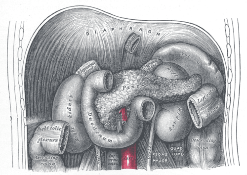
C-shaped first part of the small intestine wrapped around the head of the pancreas. It receives bile and pancreatic juice at the major duodenal papilla and is the most common site of peptic ulcers.
Core anatomy
- Location
Mostly retroperitoneal, curving around the head of the pancreas at about L1–L3.
- Parts
- 1st (superior) part (about L1): the proximal 2 cm (duodenal cap/bulb) is intraperitoneal and mobile. Most duodenal ulcers occur here
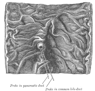
- 2nd (descending) part (L1–L3): retroperitoneal. The major duodenal papilla receives the hepatopancreatic ampulla (common bile duct + main pancreatic duct); the minor papilla receives the accessory pancreatic duct
- 3rd (horizontal) part (about L3): crosses anterior to the IVC and aorta, with the superior mesenteric vessels passing anterior to it
- 4th (ascending) part (L2–L3): ends at the duodenojejunal flexure, suspended by the suspensory muscle (ligament of Treitz)
- Relations
1st part: gallbladder neck and liver anteriorly; gastroduodenal artery, common bile duct and portal vein posteriorly. 2nd part: right kidney posteriorly, transverse colon anteriorly. 3rd part: between the superior mesenteric artery in front and the abdominal aorta behind.
- Arterial supply
The foregut–midgut junction lies just distal to the major papilla. Proximally: superior pancreaticoduodenal arteries (from the gastroduodenal, celiac trunk territory) and the supraduodenal artery. Distally: inferior pancreaticoduodenal arteries (from the superior mesenteric artery). The two form arcades around the pancreatic head.
- Venous drainage
Pancreaticoduodenal veins → superior mesenteric and portal veins.
- Innervation
Vagus and sympathetic (celiac and superior mesenteric plexuses). Visceral pain is epigastric.
Mentioned in
- Abdominal aorta
- Gallbladder and biliary tree
- Greater omentum
- Hepatic portal vein
- Inferior vena cava
- Lesser sac
- Pancreas
Generated automatically from links in other entries.


Clinical correlations
Duodenal ulcer PANCE
- Mechanism / lesion
H. pylori and NSAIDs, with acid hypersecretion. Most are in the 1st part.
- Classic presentation
Epigastric pain that improves with meals and wakes the patient at night. Posterior ulcers can erode the gastroduodenal artery (major upper GI bleed). Anterior ulcers tend to perforate, causing free air and peritonitis.
Duodenal atresia PANCE
- Mechanism / lesion
Failure of recanalization of the duodenal lumen. Strongly associated with Down syndrome (trisomy 21).
- Classic presentation
Bilious vomiting in the first day(s) of life (the obstruction is usually distal to the papilla), polyhydramnios prenatally, and a "double bubble" sign on radiograph.
Superior mesenteric artery syndrome
- Mechanism / lesion
Compression of the 3rd part between the SMA and aorta when the aortomesenteric angle narrows, typically after rapid weight loss (loss of the mesenteric fat pad) or spinal surgery.
- Classic presentation
Postprandial epigastric pain, early satiety, bilious vomiting, weight loss.
Upper vs lower GI bleeding PANCE
- Mechanism / lesion
The ligament of Treitz (duodenojejunal flexure) is the conventional dividing point.
- Classic presentation
Bleeding proximal to it is "upper GI" (hematemesis, melena, elevated BUN/creatinine ratio); distal to it is "lower GI" (typically hematochezia).
High-yield PANCE pearls
- Posterior duodenal ulcer → gastroduodenal artery hemorrhage; anterior ulcer → perforation.
- Double bubble + bilious vomiting in a newborn with Down syndrome → duodenal atresia.
- Ligament of Treitz divides upper from lower GI bleeding.
- Major duodenal papilla (2nd part) = CBD + main pancreatic duct → gallstone pancreatitis if a stone impacts here.
Imaging anatomy
- X-ray
Newborn abdominal film showing the double bubble (gas in the stomach and proximal duodenum) with little distal gas suggests duodenal atresia. Free air with a perforated ulcer.
- CT
Duodenal wall thickening, perforation (extraluminal air near the duodenum), SMA syndrome (narrow aortomesenteric angle and distance with a distended proximal duodenum).
- Ultrasound
Prenatal ultrasound can show the double bubble and polyhydramnios.
- Key views & landmarks
- EGD for ulcers and bleeding (diagnostic and therapeutic)
- Upper GI contrast series: SMA syndrome, malrotation (abnormal position of the duodenojejunal flexure)
Physical exam
- Surface landmarks
The 1st part lies near the transpyloric plane (L1) just right of the midline; the 3rd part crosses the midline at about L3.
- Palpation
Epigastric tenderness in ulcer disease. A rigid abdomen with perforation.
Self-quiz
A 52-year-old man taking daily ibuprofen has melena and hypotension. Endoscopy shows an ulcer on the posterior wall of the duodenal bulb with a visible vessel. Which artery is most likely eroded?
Correct answer: B
The gastroduodenal artery runs directly behind the 1st part of the duodenum and is the classic source of bleeding from a posterior duodenal ulcer.
Show answer
B
The gastroduodenal artery runs directly behind the 1st part of the duodenum and is the classic source of bleeding from a posterior duodenal ulcer.
A newborn with trisomy 21 has bilious vomiting on the first day of life. The radiograph shows a "double bubble." What is the diagnosis?
Correct answer: B
Duodenal atresia (failure of recanalization) is associated with Down syndrome and shows a double bubble. Pyloric stenosis presents at 3–6 weeks with non-bilious vomiting.
Show answer
B
Duodenal atresia (failure of recanalization) is associated with Down syndrome and shows a double bubble. Pyloric stenosis presents at 3–6 weeks with non-bilious vomiting.
Which structure passes anterior to the third part of the duodenum?
Correct answer: C
The SMA and SMV pass anterior to the 3rd part; the IVC and aorta lie behind it. Narrowing of the angle between the SMA and aorta can compress the duodenum (SMA syndrome).
Show answer
C
The SMA and SMV pass anterior to the 3rd part; the IVC and aorta lie behind it. Narrowing of the angle between the SMA and aorta can compress the duodenum (SMA syndrome).
After losing 15 kg rapidly, a young woman has postprandial epigastric pain, early satiety and bilious vomiting. Which part of the duodenum is compressed, and between which structures?
Correct answer: C
In SMA syndrome, loss of the mesenteric fat pad narrows the aortomesenteric angle so the 3rd part of the duodenum is compressed between the SMA in front and the aorta behind.
Show answer
C
In SMA syndrome, loss of the mesenteric fat pad narrows the aortomesenteric angle so the 3rd part of the duodenum is compressed between the SMA in front and the aorta behind.
The duodenojejunal flexure, suspended by the ligament of Treitz, is the conventional boundary for what?
Correct answer: A
The ligament of Treitz divides upper from lower GI bleeding. The foregut–midgut junction lies just distal to the major papilla in the 2nd part, and most duodenal ulcers are in the 1st part.
Show answer
A
The ligament of Treitz divides upper from lower GI bleeding. The foregut–midgut junction lies just distal to the major papilla in the 2nd part, and most duodenal ulcers are in the 1st part.