Subscapularis
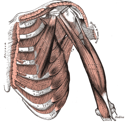
Large rotator cuff muscle on the costal surface of the scapula. It is the main medial rotator of the arm and the only cuff muscle inserting on the lesser tubercle.
Core anatomy
- Location
Subscapular fossa, forming most of the posterior wall of the axilla.
- Group
Rotator cuff (SITS); scapulohumeral muscles.
- Origin
Subscapular fossa (costal/anterior surface) of the scapula.
- Insertion
Lesser tubercle of the humerus.
- Innervation
Upper and lower subscapular nerves from the posterior cord (C5, C6) verify
- Blood supply
Subscapular artery and its branches verify
- Action
- Medially (internally) rotates and adducts the arm
- With the other rotator cuff muscles, holds the humeral head in the glenoid cavity (anterior stabilizer)
- Relations
The tendon passes anterior to the glenohumeral joint capsule, separated from the scapular neck by the subtendinous bursa of subscapularis, which usually communicates with the joint cavity. It forms part of the stabilizing sling for the long head of biceps brachii at the entrance of the intertubercular groove.
Forms a boundary of
Mentioned in
Generated automatically from links in other entries.
Swipe sideways to see the whole diagram →

Clinical correlations
Subscapularis tear
- Mechanism / lesion
Forced external rotation or hyperextension, or anterior dislocation (especially in patients over ~40). Can also be degenerative.
- Classic presentation
Anterior shoulder pain and weak internal rotation. Increased passive external rotation compared with the other side. Positive lift-off and belly-press tests. May be associated with medial subluxation/dislocation of the long head of biceps tendon.
High-yield PANCE pearls
- Subscapularis = main internal rotator; inserts on the lesser tubercle.
- Tests: lift-off (Gerber), belly-press, bear-hug.
- Consider a subscapularis tear in an older patient with persistent weakness after an anterior dislocation.
Imaging anatomy
- X-ray
Usually normal.
- MRI
Axial images are best for the subscapularis tendon and lesser tuberosity insertion. Look for an empty or medially displaced biceps tendon from its groove when the subscapularis tendon is torn.
- Ultrasound
Scanned anteriorly with the arm externally rotated. The tendon is seen in long axis inserting on the lesser tuberosity. Dynamic rotation can show medial subluxation of the biceps tendon.
- Key views & landmarks
- MRI axial proton density fat-sat through the lesser tuberosity
Physical exam
- Surface landmarks
The muscle is largely inaccessible. The tendon insertion can be palpated at the lesser tubercle, medial to the intertubercular groove, with the arm externally rotated.
- Strength / function testing
Resisted internal rotation with the elbow at the side.
Lift-off test (Gerber)
- Technique
The patient places the dorsum of the hand on the lower back (lumbar region) and tries to lift the hand away from the back.
- Positive test
Unable to lift the hand off the back, or does so only by extending the elbow or shoulder.
- Significance
Subscapularis tear or dysfunction. Requires enough internal rotation to reach the position.
Belly-press test
- Technique
The patient presses the palm against the abdomen with the wrist straight, bringing the elbow forward in front of the trunk.
- Positive test
The elbow drops back behind the trunk, or the wrist flexes to generate pressure.
- Significance
Subscapularis tear. Useful when the patient can't reach behind the back.
Self-quiz
A 62-year-old man dislocated his shoulder anteriorly 6 weeks ago. He still has anterior shoulder pain. When he places the back of his hand against his lower back, he can't lift it away. Which tendon is most likely torn?
Correct answer: D
A positive lift-off test indicates subscapularis weakness. Subscapularis is an anterior stabilizer and can tear with anterior dislocation, especially in older patients.
Show answer
D
A positive lift-off test indicates subscapularis weakness. Subscapularis is an anterior stabilizer and can tear with anterior dislocation, especially in older patients.
Subscapularis inserts onto which structure?
Correct answer: B
Subscapularis is the only rotator cuff muscle that inserts on the lesser tubercle. The other three insert on the greater tubercle.
Show answer
B
Subscapularis is the only rotator cuff muscle that inserts on the lesser tubercle. The other three insert on the greater tubercle.
Which nerves supply subscapularis?
Correct answer: B
The upper and lower subscapular nerves (posterior cord) supply subscapularis. The lower subscapular nerve also supplies teres major, and the thoracodorsal nerve supplies latissimus dorsi.
Show answer
B
The upper and lower subscapular nerves (posterior cord) supply subscapularis. The lower subscapular nerve also supplies teres major, and the thoracodorsal nerve supplies latissimus dorsi.
Ultrasound of a patient with a complete subscapularis tear shows the long head of biceps tendon out of the intertubercular groove. In which direction is it usually displaced?
Correct answer: A
The subscapularis tendon forms part of the sling that holds the biceps tendon at the entrance of the groove. When it tears, the long head of biceps can subluxate or dislocate medially.
Show answer
A
The subscapularis tendon forms part of the sling that holds the biceps tendon at the entrance of the groove. When it tears, the long head of biceps can subluxate or dislocate medially.
Which exam finding suggests a subscapularis tear?
Correct answer: D
With subscapularis (the main internal rotator) torn, the shoulder shows increased passive external rotation, weak internal rotation, and positive lift-off and belly-press tests. Reduced passive external rotation suggests adhesive capsulitis.
Show answer
D
With subscapularis (the main internal rotator) torn, the shoulder shows increased passive external rotation, weak internal rotation, and positive lift-off and belly-press tests. Reduced passive external rotation suggests adhesive capsulitis.