Muscles of mastication
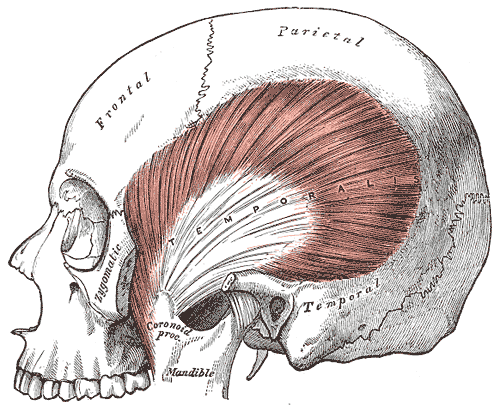
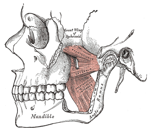
Four muscles derived from the first pharyngeal arch and supplied by the mandibular nerve (V3) — masseter, temporalis and medial pterygoid close the jaw; lateral pterygoid opens and protrudes it.
Core anatomy
- Location
Around the ramus of the mandible and the temporal fossa.
- Group
First pharyngeal arch derivatives
- Origin
Masseter: zygomatic arch. Temporalis: temporal fossa and fascia. Medial pterygoid: medial surface of the lateral pterygoid plate (deep head) and maxillary tuberosity. Lateral pterygoid: greater wing of the sphenoid (upper head) and lateral surface of the lateral pterygoid plate (lower head).
- Insertion
Masseter: lateral surface of the ramus and angle. Temporalis: coronoid process and anterior ramus. Medial pterygoid: medial surface of the ramus and angle. Lateral pterygoid: neck of the condyle, the articular disc and capsule of the temporomandibular joint.
- Innervation
Mandibular division of the trigeminal nerve (V3) — motor root
- Blood supply
Branches of the maxillary artery (from the external carotid artery).
- Action
- Elevation (closing): masseter, temporalis, medial pterygoid
- Depression (opening): lateral pterygoid (with gravity and the suprahyoid muscles)
- Protrusion: lateral and medial pterygoids
- Retraction: posterior fibers of temporalis
- Side-to-side grinding: alternating pterygoids
- Relations
Mnemonic: "M's close the Mouth" (Masseter, teMporalis, Medial pterygoid); the Lateral pterygoid Lowers (opens). The parotid gland and duct lie over the masseter.
Mentioned in
Generated automatically from links in other entries.


Clinical correlations
Trismus
- Mechanism / lesion
Spasm or inflammation of the masticatory muscles — dental infection (pericoronitis), peritonsillar or deep neck abscess, tetanus (lockjaw), radiation.
- Classic presentation
Restricted mouth opening; trismus with fever and neck swelling suggests a deep space infection.
Trigeminal (V3) motor lesion
- Mechanism / lesion
Lesion of the motor root of V3 (skull base tumors, trauma).
- Classic presentation
Weak clenching and wasting of temporalis and masseter on one side; the jaw deviates toward the weak side on opening (the intact opposite lateral pterygoid pushes it over).
Masseteric hypertrophy and bruxism
- Mechanism / lesion
Chronic clenching or grinding.
- Classic presentation
Enlarged angle of the jaw, tooth wear, TMJ pain.
High-yield PANCE pearls
- All muscles of mastication = V3.
- Lateral pterygoid opens the jaw; the others close it.
- Jaw deviates toward the side of a V3 motor lesion on opening.
- Lockjaw (trismus) → consider tetanus or deep space infection.
Imaging anatomy
- CT
Deep neck/masticator space infections and tumors.
- MRI
Masticator space masses; denervation atrophy after V3 injury.
Physical exam
- Surface landmarks
Masseter is felt at the angle of the jaw and temporalis above the zygomatic arch when the patient clenches.
- Palpation
Ask the patient to clench the teeth and palpate both masseters and temporalis muscles for bulk and symmetry.
- Strength / function testing
CN V motor exam — clenching strength, jaw opening against resistance, jaw deviation on opening; jaw-jerk reflex (brisk in upper motor neuron lesions above the pons).
Self-quiz
Which muscle of mastication opens the mouth?
Correct answer: D
The lateral pterygoid pulls the condyle and disc forward, depressing and protruding the jaw. Masseter, temporalis and medial pterygoid elevate it.
Show answer
D
The lateral pterygoid pulls the condyle and disc forward, depressing and protruding the jaw. Masseter, temporalis and medial pterygoid elevate it.
A patient's jaw deviates to the right when she opens her mouth. Which nerve is most likely injured?
Correct answer: B
The jaw deviates toward the weak side because the intact left lateral pterygoid pushes it across. So the right V3 motor supply is affected. (The tongue also deviates toward the weak side in a hypoglossal lesion.)
Show answer
B
The jaw deviates toward the weak side because the intact left lateral pterygoid pushes it across. So the right V3 motor supply is affected. (The tongue also deviates toward the weak side in a hypoglossal lesion.)
The muscles of mastication develop from which pharyngeal arch?
Correct answer: B
The muscles of mastication derive from the first pharyngeal arch and are supplied by its nerve, V3. The muscles of facial expression derive from the second arch (CN VII).
Show answer
B
The muscles of mastication derive from the first pharyngeal arch and are supplied by its nerve, V3. The muscles of facial expression derive from the second arch (CN VII).
Ten days after a puncture wound, an unvaccinated farmer can't open his mouth and has painful muscle spasms. What is the most likely diagnosis?
Correct answer: C
Tetanus causes trismus ("lockjaw") and generalized spasms. Trismus with fever and neck swelling suggests a deep space infection or peritonsillar abscess instead.
Show answer
C
Tetanus causes trismus ("lockjaw") and generalized spasms. Trismus with fever and neck swelling suggests a deep space infection or peritonsillar abscess instead.
The lateral pterygoid inserts on which structures?
Correct answer: A
The lateral pterygoid inserts on the condylar neck, articular disc and capsule of the TMJ and pulls them forward to open and protrude the jaw. Temporalis inserts on the coronoid process; masseter and medial pterygoid on the ramus and angle.
Show answer
A
The lateral pterygoid inserts on the condylar neck, articular disc and capsule of the TMJ and pulls them forward to open and protrude the jaw. Temporalis inserts on the coronoid process; masseter and medial pterygoid on the ramus and angle.