Peritoneal cavity
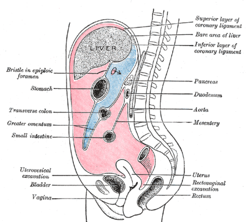
Potential space between the parietal and visceral peritoneum. Its recesses are where fluid, blood and pus collect, which is the anatomic basis of the FAST exam.
Core anatomy
- Location
Within the abdominopelvic cavity, lined by parietal peritoneum and containing the intraperitoneal organs (which are covered by visceral peritoneum).
- Boundaries
- parietal peritoneum
- Lines the abdominal and pelvic walls. Somatic innervation (lower intercostal, subcostal, iliohypogastric, ilioinguinal and phrenic nerves), so pain is sharp and localized
- visceral peritoneum
- Covers the organs. Autonomic innervation; pain is dull, poorly localized, and referred by embryologic segment
- Contents
- A small volume of serous peritoneal fluid
- Greater sac (main part) and lesser sac (omental bursa), connected through the omental (epiploic) foramen
- Divided by the transverse mesocolon into supracolic and infracolic compartments
- Communications
Paracolic gutters lateral to the ascending and descending colon connect the upper abdomen with the pelvis (fluid and infection spread along them). In females, the peritoneal cavity communicates with the exterior via the uterine tubes, uterus and vagina.
- Notes
Intraperitoneal vs retroperitoneal. Retroperitoneal organs are remembered as SAD PUCKER: Suprarenal glands, Aorta and IVC, Duodenum (2nd–4th parts), Pancreas (except the tail), Ureters, Colon (ascending and descending), Kidneys, Esophagus (abdominal part), Rectum (lower part). Key recesses: the hepatorenal recess (Morison pouch) is the most dependent part of the upper abdominal cavity when supine; the rectouterine pouch (of Douglas) in females or rectovesical pouch in males is the most dependent part when upright.
Swipe sideways to see the whole diagram →

Clinical correlations
Peritonitis PANCE
- Mechanism / lesion
Inflammation of the peritoneum from perforation (ulcer, appendix, diverticulum), infection, blood or bile.
- Classic presentation
Severe pain worsened by movement, so the patient lies still. Guarding, rigidity (board-like abdomen), rebound tenderness, percussion tenderness, fever, and absent bowel sounds (ileus).
Ascites and spontaneous bacterial peritonitis PANCE
- Mechanism / lesion
Fluid accumulates in the peritoneal cavity, most often from cirrhosis with portal hypertension. SBP is infection of ascitic fluid without a surgical source.
- Classic presentation
Abdominal distension, bulging flanks, shifting dullness, fluid wave. SBP causes fever, abdominal tenderness or encephalopathy and is diagnosed by ascitic fluid PMN count of 250 cells/mm³ or more. A serum–ascites albumin gradient (SAAG) of 1.1 g/dL or more indicates portal hypertension.
Hemoperitoneum PANCE
- Mechanism / lesion
Bleeding into the peritoneal cavity (splenic or hepatic injury, ruptured ectopic pregnancy, ruptured AAA with intraperitoneal extension).
- Classic presentation
Abdominal pain and distension, shock, and referred shoulder pain (Kehr sign). Detected at the bedside by the FAST exam.
High-yield PANCE pearls
- Morison pouch (hepatorenal recess): first place free fluid appears on supine FAST.
- SBP: ascitic PMN ≥ 250/mm³. SAAG ≥ 1.1 = portal hypertension.
- Peritonitis: rigidity, rebound, guarding; patient lies still.
- Retroperitoneal organs: SAD PUCKER.
- Upright CXR free air under the diaphragm → perforated viscus.
Imaging anatomy
- X-ray
Upright chest radiograph: free air under the diaphragm (pneumoperitoneum). Left lateral decubitus film if the patient can't stand. Supine signs of free air include the Rigler (double-wall) sign.
- CT
Most sensitive for small amounts of free air and fluid, abscesses, and the source of peritonitis.
- Ultrasound
FAST (Focused Assessment with Sonography in Trauma) views: RUQ (Morison pouch, hepatorenal), LUQ (splenorenal recess and around the spleen), pelvic (rectovesical or rectouterine pouch), and subxiphoid (pericardium); eFAST adds the pleural spaces. Free fluid appears anechoic (black). Ultrasound also guides paracentesis.
- Key views & landmarks
- FAST: RUQ, LUQ, pelvic, subxiphoid (± bilateral chest)
- Upright CXR for free air
Physical exam
- Surface landmarks
Paracentesis is performed in a lower quadrant lateral to the rectus sheath (commonly the left) or in the midline below the umbilicus, ideally ultrasound-guided.
- Palpation
Palpate gently for guarding and rigidity. Test for rebound tenderness cautiously (percussion tenderness is a gentler alternative).
- Strength / function testing
Shifting dullness: percuss the tympany–dullness border supine, then roll the patient to one side; the border shifts with ascites. Fluid wave: tap one flank while an assistant presses the midline; feel the impulse on the other side. Both are more reliable with larger volumes.
Rebound tenderness
- Technique
Press slowly and deeply over the abdomen, then release quickly.
- Positive test
Pain is worse on release than on pressure.
- Significance
Peritoneal irritation.
Self-quiz
A 28-year-old man is hypotensive after a motorcycle crash. Which location is the most dependent site in the upper abdomen where free fluid is typically first seen on a supine FAST exam?
Correct answer: B
In the supine patient the hepatorenal recess (Morison pouch) is the most dependent part of the upper peritoneal cavity, so the RUQ view is often positive first. The pelvis is most dependent when upright.
Show answer
B
In the supine patient the hepatorenal recess (Morison pouch) is the most dependent part of the upper peritoneal cavity, so the RUQ view is often positive first. The pelvis is most dependent when upright.
A patient with cirrhosis and ascites has fever and diffuse abdominal tenderness. Diagnostic paracentesis shows 480 neutrophils/mm³. What is the diagnosis?
Correct answer: B
An ascitic PMN count of 250/mm³ or more diagnoses SBP. Treat with empiric antibiotics (e.g. a third-generation cephalosporin) and albumin in selected patients.
Show answer
B
An ascitic PMN count of 250/mm³ or more diagnoses SBP. Treat with empiric antibiotics (e.g. a third-generation cephalosporin) and albumin in selected patients.
Which organ is retroperitoneal?
Correct answer: C
The 2nd–4th parts of the duodenum are retroperitoneal (SAD PUCKER). The stomach, transverse colon, spleen and sigmoid colon are intraperitoneal.
Show answer
C
The 2nd–4th parts of the duodenum are retroperitoneal (SAD PUCKER). The stomach, transverse colon, spleen and sigmoid colon are intraperitoneal.
In an upright woman, which is the most dependent part of the peritoneal cavity?
Correct answer: C
The rectouterine pouch (rectovesical pouch in males) is the lowest point when upright, so fluid collects there. The hepatorenal recess (Morison pouch) is the most dependent part of the upper abdomen when supine.
Show answer
C
The rectouterine pouch (rectovesical pouch in males) is the lowest point when upright, so fluid collects there. The hepatorenal recess (Morison pouch) is the most dependent part of the upper abdomen when supine.
A serum–ascites albumin gradient (SAAG) of 1.1 g/dL or more indicates what?
Correct answer: A
A SAAG ≥ 1.1 g/dL indicates portal hypertension (most often cirrhosis). A low SAAG suggests peritoneal causes such as carcinomatosis, TB or pancreatitis. SBP is diagnosed by an ascitic PMN count of 250/mm³ or more.
Show answer
A
A SAAG ≥ 1.1 g/dL indicates portal hypertension (most often cirrhosis). A low SAAG suggests peritoneal causes such as carcinomatosis, TB or pancreatitis. SBP is diagnosed by an ascitic PMN count of 250/mm³ or more.