Sacroiliac joint
Strong, nearly immobile joint between the sacrum and ilium that transmits body weight to the pelvis. It is the classic site of inflammatory sacroiliitis in axial spondyloarthritis.
Core anatomy
- Location
Between the auricular surfaces of the sacrum and coccyx and the ilium, deep to the PSIS on each side.
- Classification
Compound joint: a synovial (plane) joint anteriorly between the auricular surfaces, and a syndesmosis posteriorly (interosseous ligament between the tuberosities).
- Articulating surfaces
Sacrum and coccyx and the ilium of the hip bone
- Capsule
Thin anteriorly; the articular surfaces are irregular and interlocking, with hyaline cartilage (thicker on the sacral side) and fibrocartilage.
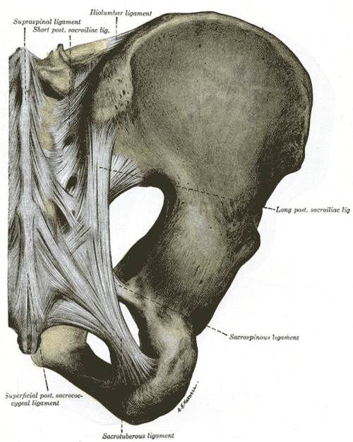
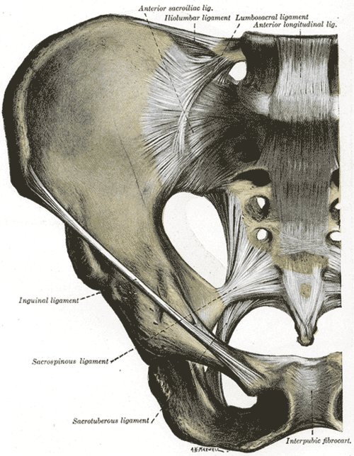
- Ligaments
- Interosseous sacroiliac ligament: the strongest; fills the space between the sacral and iliac tuberosities
- Posterior sacroiliac ligaments: posterior to the interosseous ligament
- Anterior sacroiliac ligament: thin, part of the anterior capsule
- Iliolumbar ligament: L5 transverse process to the iliac crest
- Accessory: sacrotuberous and sacrospinous ligaments, which resist upward tilting of the lower sacrum
- Movements
- Very limited gliding and rotation (nutation and counternutation of the sacrum), mainly during pregnancy and changes of posture
- Stability
Body weight tends to drive the sacrum downward and rotate its upper end forward; the interlocking surfaces, the interosseous and posterior ligaments and the sacrotuberous/sacrospinous ligaments resist this. Relaxin in pregnancy loosens the ligaments.
- Innervation
Mainly lateral branches of the dorsal rami of about L5–S3 posteriorly, with anterior contributions from ventral rami verify
- Blood supply
Iliolumbar, superior gluteal and lateral sacral arteries (branches of the internal iliac artery).
- Relations
Anterior: the psoas and the lumbosacral trunk, obturator nerve and internal iliac vessels cross the pelvic brim nearby; the ureter crosses the pelvic brim just anterior to the joint. Posterior: erector spinae and gluteus maximus.
Mentioned in
Generated automatically from links in other entries.


Clinical correlations
Sacroiliitis in axial spondyloarthritis (ankylosing spondylitis) PANCE
- Mechanism / lesion
Immune-mediated enthesitis and synovitis of the SI joints, strongly associated with HLA-B27. Also seen with IBD-associated, psoriatic and reactive arthritis (these are more often asymmetric).
- Classic presentation
Inflammatory back pain in a young adult (onset before about 40): insidious, lasting over 3 months, morning stiffness over 30 minutes, night pain, better with exercise and not with rest; alternating buttock pain. Extra-articular: anterior uveitis, enthesitis (Achilles), dactylitis, reduced chest expansion.
- Notes
First-line treatment is NSAIDs plus exercise and physical therapy; TNF inhibitors (or IL-17 inhibitors) if symptoms persist. Methotrexate does not help axial disease.
Sacroiliac joint dysfunction
- Mechanism / lesion
Mechanical pain from the joint or its ligaments, e.g. after pregnancy, falls onto the buttock, or leg-length discrepancy.
- Classic presentation
Unilateral pain below L5 over the PSIS (the patient points to it with one finger — the Fortin finger test), sometimes radiating to the buttock or posterior thigh but not below the knee.
Septic sacroiliitis
- Mechanism / lesion
Hematogenous infection (Staphylococcus aureus most often), with risk factors such as injection drug use.
- Classic presentation
Fever, buttock or low back pain, limp; easily mistaken for a disc or hip problem. MRI is the key test.
High-yield PANCE pearls
- Young man, chronic low back pain with morning stiffness better with exercise, bilateral sacroiliitis → ankylosing spondylitis (HLA-B27).
- AS: first-line NSAIDs; then TNF inhibitors.
- AS extra-articular association to remember: anterior uveitis.
- MRI (STIR) detects early sacroiliitis before X-ray changes.
- FABER with posterior (SI) pain vs groin pain (hip joint).
Imaging anatomy
- X-ray
AP pelvis or angled AP (Ferguson view) of the SI joints: erosions, "pseudo-widening", sclerosis, then ankylosis, usually bilateral and symmetric in AS. Changes may take years to appear. Late spine: squaring of vertebral bodies, syndesmophytes, "bamboo spine".
- CT
Best for bony erosions and ankylosis; also used to guide SI joint injections.
- MRI
STIR/T2 fat-suppressed MRI shows bone marrow edema (active sacroiliitis) before radiographic damage, and is the test of choice for suspected early axial spondyloarthritis or septic sacroiliitis.
- Key views & landmarks
- AP pelvis or Ferguson view of the SI joints
- MRI of the SI joints (STIR)
Physical exam
- Surface landmarks
The SI joint lies deep to the PSIS (the "dimples of Venus", at about S2).
- Palpation
Tenderness over the sacral sulcus just medial to the PSIS.
- Strength / function testing
Schober test for lumbar flexion in suspected AS: mark the skin at the level of the dimples of Venus and 10 cm above; an increase of less than 5 cm on full forward flexion suggests restricted lumbar motion. Chest expansion is also reduced in advanced AS.
FABER (Patrick) test
- Technique
Supine; the tested leg is placed in Flexion, ABduction and External Rotation (figure-4, heel on the opposite knee). The examiner presses the knee down while stabilizing the opposite ASIS.
- Positive test
Pain in the posterior pelvis/SI region (SI joint), or in the groin (hip joint).
- Significance
Screens for SI joint and hip pathology; not specific.
Gaenslen test
- Technique
Supine at the edge of the table; one hip is fully flexed toward the chest while the other leg is lowered over the edge into hyperextension.
- Positive test
Pain in the SI joint.
- Significance
SI joint provocation test.
Thigh thrust (posterior shear) test
- Technique
Supine with the hip flexed to 90°; the examiner applies an axial force along the femur toward the table while supporting the sacrum.
- Positive test
Reproduction of the patient's buttock pain.
- Significance
One of the more sensitive single SI provocation tests. Several positive provocation tests together make SI joint pain more likely. verify
Self-quiz
A 24-year-old man has 8 months of low back and alternating buttock pain. He is stiff for over an hour each morning and feels better after exercise. He once had a painful red eye with photophobia. Radiographs are normal. What is the best next imaging study?
Correct answer: B
This is inflammatory back pain with a history suggesting anterior uveitis, pointing to axial spondyloarthritis. Radiographic sacroiliitis can lag for years; MRI of the SI joints shows active bone marrow edema early (non-radiographic axial SpA).
Show answer
B
This is inflammatory back pain with a history suggesting anterior uveitis, pointing to axial spondyloarthritis. Radiographic sacroiliitis can lag for years; MRI of the SI joints shows active bone marrow edema early (non-radiographic axial SpA).
What is the first-line drug treatment for ankylosing spondylitis?
Correct answer: C
NSAIDs (with exercise and physical therapy) are first-line and can reduce inflammatory pain. Persistent disease is treated with TNF inhibitors or IL-17 inhibitors. Conventional DMARDs such as methotrexate and sulfasalazine do not help axial disease, and systemic steroids are not used long term.
Show answer
C
NSAIDs (with exercise and physical therapy) are first-line and can reduce inflammatory pain. Persistent disease is treated with TNF inhibitors or IL-17 inhibitors. Conventional DMARDs such as methotrexate and sulfasalazine do not help axial disease, and systemic steroids are not used long term.
Which is the strongest ligament of the sacroiliac joint?
Correct answer: B
The interosseous sacroiliac ligament, which forms the posterior syndesmotic part of the joint, is the strongest and main load-bearing ligament. The anterior ligament is thin.
Show answer
B
The interosseous sacroiliac ligament, which forms the posterior syndesmotic part of the joint, is the strongest and main load-bearing ligament. The anterior ligament is thin.
How is the sacroiliac joint classified?
Correct answer: D
The sacroiliac joint is a compound joint: a synovial (plane) joint between the auricular surfaces anteriorly and a syndesmosis (interosseous ligament between the tuberosities) posteriorly. It allows only slight gliding and rotation (nutation).
Show answer
D
The sacroiliac joint is a compound joint: a synovial (plane) joint between the auricular surfaces anteriorly and a syndesmosis (interosseous ligament between the tuberosities) posteriorly. It allows only slight gliding and rotation (nutation).
A 27-year-old man with ankylosing spondylitis still has inflammatory back pain despite trials of two NSAIDs and regular exercise. What is the most appropriate next treatment?
Correct answer: B
First-line treatment for axial spondyloarthritis is NSAIDs plus exercise and physical therapy; a TNF inhibitor (or IL-17 inhibitor) is used when symptoms persist. Methotrexate does not help axial disease, and rest makes inflammatory back pain worse.
Show answer
B
First-line treatment for axial spondyloarthritis is NSAIDs plus exercise and physical therapy; a TNF inhibitor (or IL-17 inhibitor) is used when symptoms persist. Methotrexate does not help axial disease, and rest makes inflammatory back pain worse.