Bones of the foot
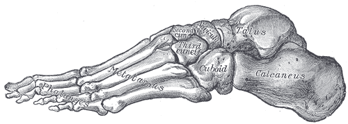
Seven tarsals, five metatarsals and fourteen phalanges forming the arches of the foot. Fifth metatarsal fractures, calcaneal fractures, stress fractures and plantar fasciitis are the common problems.
Core anatomy
- Location
Below the ankle joint; the talus articulates with the tibia and fibula.
- Classification
Short bones (tarsals) and long bones (metatarsals, phalanges)
- Key features & landmarks
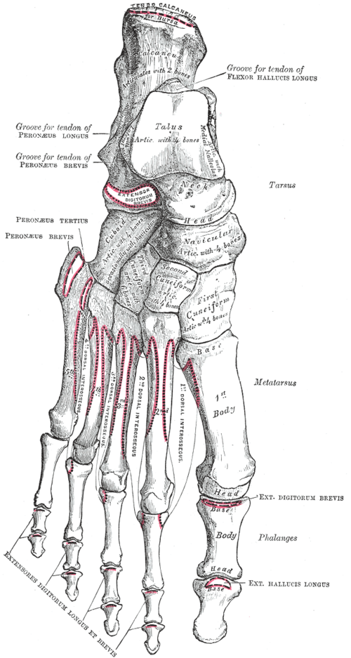
- Talus: body, neck and head; no muscle attachments; its blood supply enters mainly through the neck, so neck fractures risk avascular necrosis
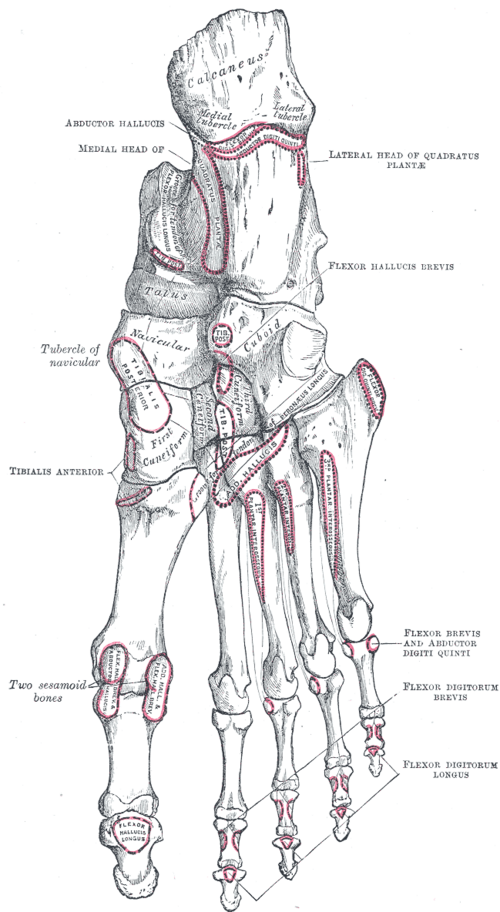
- Calcaneus: the largest tarsal (heel); the sustentaculum tali supports the talus; the calcaneal tuberosity receives the Achilles tendon and gives origin to the plantar fascia
- Navicular (with a tuberosity for tibialis posterior), cuboid (groove for fibularis longus) and three cuneiforms
- Metatarsals: the 5th metatarsal tuberosity receives fibularis brevis; the 1st metatarsal head has two sesamoids in the flexor hallucis brevis tendons
- Phalanges: two in the great toe, three in each of the others
- Joints: subtalar (inversion/eversion), transverse tarsal (Chopart), tarsometatarsal (Lisfranc)
- Arches: medial longitudinal (highest; supported by tibialis posterior, the spring ligament and plantar fascia), lateral longitudinal and transverse
- Relations
The tibial nerve and posterior tibial artery pass behind the medial malleolus through the tarsal tunnel to the sole; the dorsalis pedis artery lies on the dorsum between the extensor hallucis longus and extensor digitorum longus tendons.
- Ossification
The calcaneus and talus ossify before birth; the navicular is the last tarsal to ossify (about 3 years), which is relevant to Köhler disease (AVN of the navicular in children).
Muscle attachments
- Triceps surae and calcaneal tendon — insertion
Joints
Generated automatically from links in other entries.



Clinical correlations
Fifth metatarsal fractures PANCE
- Mechanism / lesion
Avulsion (pseudo-Jones) fracture of the tuberosity from an inversion injury (fibularis brevis pull). Jones fracture: at the metaphyseal–diaphyseal junction, in a watershed area.
- Classic presentation
Lateral midfoot pain after an inversion injury. Avulsions heal well in a walking boot; Jones fractures have a high risk of nonunion (non-weight-bearing cast or surgical fixation in athletes).
Plantar fasciitis PANCE
- Mechanism / lesion
Degenerative overload of the plantar fascia at its origin on the medial calcaneal tuberosity (running, obesity, prolonged standing, flat or high-arched feet).
- Classic presentation
Sharp heel pain with the first steps in the morning or after rest, improving with activity; tenderness at the medial calcaneal tubercle. Treat with stretching, supportive footwear, NSAIDs and night splints.
Calcaneal fracture PANCE
- Mechanism / lesion
Axial load — a fall from height onto the heels.
- Classic presentation
Heel pain and swelling, inability to bear weight; look for associated lumbar spine compression fractures and a contralateral calcaneal fracture. Böhler angle is reduced on the lateral X-ray.
Lisfranc injury
- Mechanism / lesion
Disruption of the tarsometatarsal joints (the Lisfranc ligament runs from the medial cuneiform to the 2nd metatarsal base) from an axial load on a plantarflexed foot.
- Classic presentation
Midfoot pain and swelling, plantar ecchymosis; easily missed — weight-bearing X-rays show widening between the 1st and 2nd metatarsal bases.
Metatarsal stress fracture ("march fracture") PANCE
- Mechanism / lesion
Repetitive loading, usually the 2nd (or 3rd) metatarsal shaft, in runners and recruits; consider the female athlete triad.
- Classic presentation
Gradual forefoot pain with focal tenderness; X-rays may be normal for 2–3 weeks — MRI if needed.
High-yield PANCE pearls
- Inversion injury + base of 5th metatarsal: avulsion heals well; Jones fracture → nonunion risk.
- Heel pain with the first morning steps → plantar fasciitis.
- Fall from height with calcaneal fracture → check the lumbar spine.
- Midfoot injury with plantar bruising → Lisfranc injury → weight-bearing films.
- 2nd metatarsal stress fracture = march fracture.
Imaging anatomy
- X-ray
Foot AP, oblique and lateral views; Ottawa foot rules decide who needs X-rays (tenderness at the navicular or base of the 5th metatarsal, or inability to bear weight). Weight-bearing views for Lisfranc injury.
- CT
Calcaneal and complex midfoot fractures.
- MRI
Stress fractures, AVN of the talus or navicular, occult fractures.
- Ultrasound
Thickened plantar fascia (over about 4 mm) in plantar fasciitis. verify
- Key views & landmarks
- Foot AP, oblique and lateral (weight-bearing when possible)
- Harris (axial) view of the calcaneus
Physical exam
- Surface landmarks
Base of the 5th metatarsal (lateral midfoot), navicular tuberosity (medial midfoot), medial calcaneal tubercle (plantar heel), sustentaculum tali (below the medial malleolus), head of the talus (in front of the ankle on eversion/inversion).
- Palpation
Apply the Ottawa foot rules after trauma; palpate the plantar fascia origin, metatarsal shafts and the Lisfranc joint.
- Strength / function testing
Inspect the arches standing (pes planus/cavus); single-leg heel raise tests tibialis posterior function.
Self-quiz
A 45-year-old woman has sharp pain at the bottom of her right heel that is worst with her first steps in the morning and improves after walking for a few minutes. There is tenderness at the medial calcaneal tubercle. What is the most likely diagnosis?
Correct answer: B
Plantar fasciitis causes heel pain with the first steps after rest, with tenderness at the fascia's origin on the medial calcaneal tubercle. Treatment is conservative (stretching, footwear, NSAIDs).
Show answer
B
Plantar fasciitis causes heel pain with the first steps after rest, with tenderness at the fascia's origin on the medial calcaneal tubercle. Treatment is conservative (stretching, footwear, NSAIDs).
A college basketball player has a fracture at the metaphyseal–diaphyseal junction of the 5th metatarsal. What is the main concern?
Correct answer: B
A Jones fracture occurs in a vascular watershed area and has a high rate of nonunion; athletes often have surgical fixation. Tuberosity avulsion fractures heal well with a boot.
Show answer
B
A Jones fracture occurs in a vascular watershed area and has a high rate of nonunion; athletes often have surgical fixation. Tuberosity avulsion fractures heal well with a boot.
A worker falls 4 m and lands on his feet. X-rays show a comminuted calcaneal fracture. Which other area should be imaged?
Correct answer: B
Axial load through the heels is transmitted up the skeleton; calcaneal fractures are commonly associated with lumbar (thoracolumbar) compression fractures and fractures of the opposite calcaneus.
Show answer
B
Axial load through the heels is transmitted up the skeleton; calcaneal fractures are commonly associated with lumbar (thoracolumbar) compression fractures and fractures of the opposite calcaneus.
After a fall onto a plantarflexed foot, a patient has midfoot pain, swelling and bruising on the sole. Non-weight bearing radiographs look normal. What is the best next step?
Correct answer: B
Midfoot pain with plantar ecchymosis suggests a Lisfranc (tarsometatarsal) injury, which is easily missed. Weight-bearing radiographs reveal widening between the bases of the 1st and 2nd metatarsals.
Show answer
B
Midfoot pain with plantar ecchymosis suggests a Lisfranc (tarsometatarsal) injury, which is easily missed. Weight-bearing radiographs reveal widening between the bases of the 1st and 2nd metatarsals.
Which tarsal bone has no muscle attachments and is at risk of avascular necrosis after a fracture through its neck?
Correct answer: D
The talus has no muscle attachments, and its blood supply enters mainly through the neck, so neck fractures risk AVN of the body. The calcaneus is the largest tarsal and receives the Achilles tendon.
Show answer
D
The talus has no muscle attachments, and its blood supply enters mainly through the neck, so neck fractures risk AVN of the body. The calcaneus is the largest tarsal and receives the Achilles tendon.