Sciatic nerve
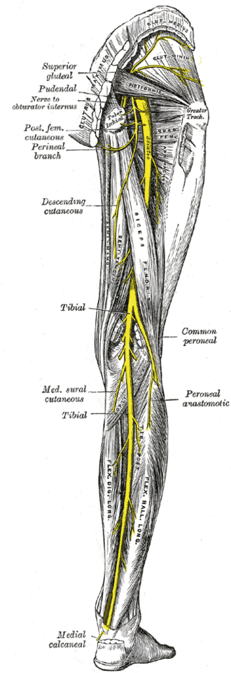
The largest nerve in the body (L4–S3), running from the buttock down the back of the thigh and dividing into the tibial and common fibular nerves. It supplies the hamstrings and everything below the knee except a strip of skin on the medial leg.
Core anatomy
- Location
Gluteal region and posterior compartment of the thigh.
- Root values
Ventral rami of L4–S3.
- Origin
Sacral plexus, on the anterior surface of piriformis
- Course
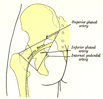
Leaves the pelvis through the greater sciatic foramen below piriformis, descends deep to gluteus maximus midway between the greater trochanter and ischial tuberosity, crosses the short lateral rotators, and runs down the posterior thigh deep to the long head of biceps femoris. It usually divides at the apex of the popliteal fossa (the division level varies, and it may divide in the pelvis).
- Branches
- Tibial nerve (L4–S3; anterior divisions)
- Common fibular nerve (L4–S2; posterior divisions)
- Muscular branches in the thigh: Hamstrings and the hamstring part of adductor magnus (tibial division); short head of biceps femoris (common fibular division)
- Motor
The hamstrings and hamstring part of adductor magnus, and — through its branches — all muscles of the leg and foot.
- Sensory
Through its branches, the skin of the leg and foot except the medial side (saphenous nerve).
- Relations
The posterior femoral cutaneous nerve lies superficial to it in the buttock; the inferior gluteal vessels accompany it.
Muscles innervated
- Adductor muscles of the thigh — obturator nerve (L2–L4) for most; pectineus mainly by the femoral nerve; the hamstring part of adductor magnus by the tibial division of the sciatic nerve
- Hamstrings — sciatic nerve: tibial division (L5–S2) for the long head of biceps, semitendinosus and semimembranosus; common fibular division for the short head of biceps femoris
Muscles innervated via branches
- Triceps surae and calcaneal tendon — via Tibial nerve
Branches
Mentioned in
Generated automatically from links in other entries.
Swipe sideways to see the whole diagram →


Clinical correlations
Sciatica (lumbosacral radiculopathy) PANCE
- Mechanism / lesion
Most often compression of the L5 or S1 nerve root by a herniated disc (see lumbar vertebrae and intervertebral disc) — not the sciatic nerve itself.
- Classic presentation
Pain radiating from the buttock down the back or side of the leg below the knee, often with paresthesias; positive straight-leg raise. Most improve with conservative care; red flags (cauda equina, progressive weakness) need urgent MRI.
Sciatic nerve injury PANCE
- Mechanism / lesion
Posterior hip dislocation and acetabular fractures, hip arthroplasty, misplaced gluteal intramuscular injections, prolonged compression (e.g. in intoxication).
- Classic presentation
Weak knee flexion and all muscles below the knee (flail foot) with sensory loss below the knee except the medial leg; ankle reflex lost. The common fibular division is more vulnerable, so partial injuries often present as foot drop.
High-yield PANCE pearls
- Sciatica is usually an L5/S1 root problem → positive straight-leg raise.
- Posterior hip dislocation → sciatic (fibular division) injury → foot drop.
- Gluteal IM injections go in the upper outer quadrant.
- Sciatic nerve divides into tibial and common fibular nerves, usually at the popliteal fossa.
Imaging anatomy
- MRI
Lumbar MRI for radiculopathy; MR neurography of the pelvis and thigh for nerve lesions, tumors and entrapment.
- Ultrasound
Shows the nerve in the thigh; guides sciatic (popliteal or subgluteal) nerve blocks.
- Key views & landmarks
- MRI lumbar spine (if red flags or persistent symptoms)
Physical exam
- Surface landmarks
The nerve enters the thigh at about the midpoint between the greater trochanter and the ischial tuberosity.
- Strength / function testing
Knee flexion, ankle dorsiflexion (deep fibular), eversion (superficial fibular), plantarflexion and inversion (tibial), great toe extension (L5); ankle reflex (S1); sensation of the sole, dorsum and lateral leg.
Straight-leg raise (Lasègue) test
- Technique
Supine; raise the extended leg passively.
- Positive test
Radiating pain below the knee between about 30° and 70° of hip flexion (thigh tightness alone doesn't count).
- Significance
Sensitive for L5/S1 radiculopathy; a crossed straight-leg raise (pain in the affected leg when the other leg is raised) is more specific.
Self-quiz
A patient received an intramuscular injection into the lower medial part of the left buttock. He now has weakness of ankle dorsiflexion and eversion with numbness on the dorsum of the foot. Which nerve fibers were most likely injured?
Correct answer: B
The sciatic nerve lies in the lower medial buttock. Its common fibular division is more vulnerable, so partial injuries cause foot drop and dorsal foot numbness. Injections belong in the upper outer quadrant.
Show answer
B
The sciatic nerve lies in the lower medial buttock. Its common fibular division is more vulnerable, so partial injuries cause foot drop and dorsal foot numbness. Injections belong in the upper outer quadrant.
A crossed straight-leg raise test is positive. What is the significance?
Correct answer: A
Pain in the affected leg when the opposite leg is raised is specific for a herniated lumbar disc (usually a large central/axillary herniation); the ordinary SLR is sensitive but less specific.
Show answer
A
Pain in the affected leg when the opposite leg is raised is specific for a herniated lumbar disc (usually a large central/axillary herniation); the ordinary SLR is sensitive but less specific.
Through which opening, and in relation to which muscle, does the sciatic nerve usually leave the pelvis?
Correct answer: A
The sciatic nerve (L4–S3) leaves the pelvis through the greater sciatic foramen below piriformis, then descends deep to gluteus maximus midway between the greater trochanter and ischial tuberosity. The superior gluteal nerve passes above piriformis.
Show answer
A
The sciatic nerve (L4–S3) leaves the pelvis through the greater sciatic foramen below piriformis, then descends deep to gluteus maximus midway between the greater trochanter and ischial tuberosity. The superior gluteal nerve passes above piriformis.
To avoid the sciatic nerve, gluteal intramuscular injections should be given in which quadrant of the buttock?
Correct answer: D
The sciatic nerve runs through the lower medial buttock, so gluteal injections go in the upper outer quadrant. Injections placed low and medially can injure the nerve, often its common fibular division.
Show answer
D
The sciatic nerve runs through the lower medial buttock, so gluteal injections go in the upper outer quadrant. Injections placed low and medially can injure the nerve, often its common fibular division.
After complete transection of the sciatic nerve in the mid-thigh, which function remains intact?
Correct answer: C
Through its branches the sciatic nerve supplies all muscles below the knee and the skin of the leg and foot except the medial side, which is supplied by the saphenous nerve (femoral). A complete lesion causes a flail foot and loss of the ankle reflex.
Show answer
C
Through its branches the sciatic nerve supplies all muscles below the knee and the skin of the leg and foot except the medial side, which is supplied by the saphenous nerve (femoral). A complete lesion causes a flail foot and loss of the ankle reflex.