Superior vena cava
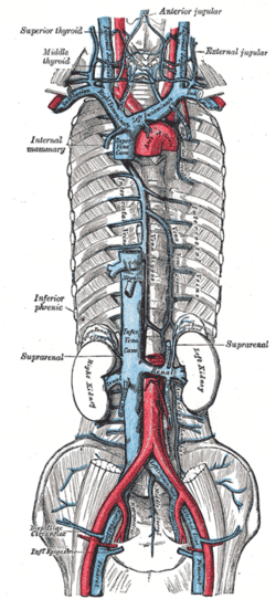
Large vein returning blood from the head, neck, upper limbs and upper thorax to the right atrium. It is the site of SVC syndrome and the target position for central venous catheter tips.
Core anatomy
- Location
Superior and middle mediastinum, to the right of the ascending aorta.
- Formation
Union of the right and left brachiocephalic veins posterior to the lower border of the right 1st costal cartilage.
- Course
Descends vertically to the right of the ascending aorta. Receives the azygos vein posteriorly at about the level of the sternal angle, just before entering the pericardium, and ends in the right atrium at about the level of the right 3rd costal cartilage. verify
- Tributaries
- Right and left brachiocephalic veins (formed by the internal jugular and subclavian veins)
- Azygos vein
- Small pericardial and mediastinal veins
- Drains into
Right atrium of the heart.
- Relations
The right phrenic nerve runs on its right side, and the right lung and pleura lie lateral to it. The ascending aorta is on its left, and the right pulmonary artery passes behind it. The SA node lies at its junction with the right atrium.
Tributaries
Found in
Mentioned in
Generated automatically from links in other entries.

Clinical correlations
Superior vena cava syndrome PANCE
- Mechanism / lesion
Obstruction of the SVC, most often by malignancy (lung cancer, especially small cell, and lymphoma), and increasingly by thrombosis related to central venous catheters or pacemaker leads.
- Classic presentation
Facial and neck swelling/plethora, upper-extremity edema, dilated collateral veins over the chest wall, dyspnea, cough, headache worse when bending forward or lying down. Pemberton sign: facial plethora on raising both arms. Severe cases have laryngeal or cerebral edema.
Central venous catheter tip position PANCE
- Mechanism / lesion
Catheters placed via the internal jugular or subclavian veins are advanced into the SVC.
- Classic presentation
The tip should lie in the lower SVC or at the cavoatrial junction. On CXR, the carina is a practical landmark (the cavoatrial junction lies below it). Always check for pneumothorax after IJ or subclavian placement. verify
Persistent left superior vena cava
- Mechanism / lesion
Failure of the left anterior cardinal vein to regress. It usually drains into the coronary sinus.
- Classic presentation
Usually asymptomatic. Seen as a left-sided catheter course on CXR or a dilated coronary sinus on echo.
High-yield PANCE pearls
- SVC syndrome: facial swelling, arm edema, chest wall collaterals; think lung cancer (small cell) or lymphoma.
- Central line tip: lower SVC/cavoatrial junction; get a CXR for position and pneumothorax.
- SVC is formed by the right + left brachiocephalic veins behind the right 1st costal cartilage.
Imaging anatomy
- X-ray
Widening of the right superior mediastinum (mass). Catheter tip position.
- CT
Contrast CT is the key test for SVC syndrome: shows the cause (mass, thrombus) and the collateral pathways (azygos, internal thoracic, chest wall veins).
- Ultrasound
Ultrasound guides internal jugular and subclavian cannulation. Upper-extremity duplex detects associated thrombosis.
- Key views & landmarks
- CT chest with IV contrast (venous phase)
- CXR after central line placement
Physical exam
- Surface landmarks
Lies behind the right border of the sternum from the 1st to the 3rd costal cartilages.
- Palpation
Distended, non-pulsatile neck veins in SVC obstruction (unlike the pulsatile JVP of right heart failure).
- Strength / function testing
Inspect for facial plethora, arm swelling and dilated chest wall veins. Pemberton maneuver: raise both arms above the head for about a minute and watch for facial plethora, cyanosis or respiratory distress.
Self-quiz
A 64-year-old man with a 60-pack-year smoking history has facial swelling, arm edema and dilated veins over his upper chest. His face becomes plethoric when he raises both arms. What is the most likely underlying cause?
Correct answer: B
SVC syndrome (positive Pemberton sign) in a heavy smoker is most likely caused by a lung cancer, classically small cell, compressing or invading the SVC. Lymphoma is the other major malignant cause.
Show answer
B
SVC syndrome (positive Pemberton sign) in a heavy smoker is most likely caused by a lung cancer, classically small cell, compressing or invading the SVC. Lymphoma is the other major malignant cause.
Where should the tip of a central venous catheter placed through the right internal jugular vein ideally lie?
Correct answer: C
The tip should lie in the lower SVC or at the cavoatrial junction. Tips in the right ventricle can cause arrhythmias, and tips in small veins increase the risk of thrombosis and perforation.
Show answer
C
The tip should lie in the lower SVC or at the cavoatrial junction. Tips in the right ventricle can cause arrhythmias, and tips in small veins increase the risk of thrombosis and perforation.
Which vein enters the posterior aspect of the SVC just above the pericardium?
Correct answer: B
The azygos vein arches over the right lung root at about the level of the sternal angle (T4) and drains into the posterior SVC.
Show answer
B
The azygos vein arches over the right lung root at about the level of the sternal angle (T4) and drains into the posterior SVC.
A persistent left superior vena cava usually drains into which structure?
Correct answer: E
A persistent left SVC (failure of the left anterior cardinal vein to regress) usually drains into the coronary sinus, which appears dilated on echocardiography. It is usually asymptomatic and may be found as a left-sided catheter course.
Show answer
E
A persistent left SVC (failure of the left anterior cardinal vein to regress) usually drains into the coronary sinus, which appears dilated on echocardiography. It is usually asymptomatic and may be found as a left-sided catheter course.
How and where is the superior vena cava formed?
Correct answer: B
The SVC is formed by the right and left brachiocephalic veins behind the lower border of the right 1st costal cartilage. It receives the azygos vein posteriorly and ends in the right atrium.
Show answer
B
The SVC is formed by the right and left brachiocephalic veins behind the lower border of the right 1st costal cartilage. It receives the azygos vein posteriorly and ends in the right atrium.