Accessory nerve (CN XI)
Motor nerve to sternocleidomastoid and trapezius, arising from the upper cervical cord. It crosses the posterior triangle superficially, where lymph node biopsy can injure it and cause shoulder droop.
Core anatomy
- Origin
Spinal accessory nucleus in the ventral horn of the upper cervical cord (about C1–C5). The rootlets ascend beside the spinal cord through the foramen magnum into the skull. (A small "cranial root" from the nucleus ambiguus joins the vagus and is usually considered part of CN X.)
- Course
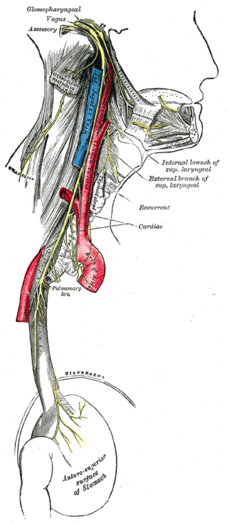
Exits through the jugular foramen with CN IX and X → descends, often crossing in front of or behind the internal jugular vein → pierces and supplies sternocleidomastoid → crosses the posterior triangle superficially on levator scapulae, just deep to the investing fascia (emerging about a third of the way down the posterior border of SCM, above the nerve point) → supplies trapezius from its deep surface (see triangles of the neck).
- Motor
Sternocleidomastoid (turns the face to the opposite side) and trapezius (shrugs, elevates and retracts the scapula, rotates it upward for abduction above 90°). C2–C4 fibers from the cervical plexus add proprioception.
- Relations
In the posterior triangle it lies superficially among the occipital and posterior cervical (level V) lymph nodes.
Muscles innervated
- Sternocleidomastoid — Spinal accessory nerve (CN XI) (motor); C2–C3 ventral rami (proprioception, via the cervical plexus).
- Trapezius — Spinal accessory nerve (CN XI) for motor; C3–C4 (cervical plexus) for pain and proprioception.
Mentioned in
Generated automatically from links in other entries.
Swipe sideways to see the whole diagram →

Clinical correlations
Spinal accessory nerve injury PANCE
- Mechanism / lesion
Iatrogenic — posterior triangle lymph node biopsy, neck dissection; also penetrating trauma.
- Classic presentation
Injury in the posterior triangle spares SCM (supplied higher up) but paralyzes trapezius: shoulder droop, weak shrug, difficulty abducting above 90°, lateral scapular winging (the inferior angle moves laterally — contrast with the medial winging of a long thoracic nerve palsy), shoulder pain.
Proximal (jugular foramen) lesions
- Mechanism / lesion
Tumors at the jugular foramen or skull base (with CN IX and X palsies).
- Classic presentation
Weak SCM (weak head turning to the opposite side) and trapezius on the same side as the lesion.
High-yield PANCE pearls
- Shoulder droop and weak shrug after a posterior triangle node biopsy → CN XI injury.
- Trapezius palsy → lateral scapular winging; long thoracic nerve (serratus anterior) → medial winging.
- Weak head turning to the left → right SCM / right CN XI.
Imaging anatomy
- MRI
MRI of the skull base and neck for unexplained XI palsy; EMG/nerve conduction confirms denervation of trapezius.
Physical exam
- Strength / function testing
SCM: turn the head against resistance (turning to the left tests the right SCM) and look/palpate for contraction. Trapezius: shrug the shoulders against resistance; look for asymmetry, wasting and scapular position; abduct the arms above 90°.
Self-quiz
Three weeks after excision of a lymph node from the right posterior triangle, a patient cannot shrug his right shoulder fully and has trouble lifting his arm above his head. Which muscle is weak?
Correct answer: B
The spinal accessory nerve crosses the posterior triangle superficially and is at risk during node biopsy. Trapezius weakness causes shoulder droop, weak shrug and difficulty abducting above 90°.
Show answer
B
The spinal accessory nerve crosses the posterior triangle superficially and is at risk during node biopsy. Trapezius weakness causes shoulder droop, weak shrug and difficulty abducting above 90°.
The spinal accessory nerve arises from which part of the nervous system?
Correct answer: B
Its motor neurons lie in the ventral horn of C1–C5; the rootlets ascend through the foramen magnum and leave through the jugular foramen.
Show answer
B
Its motor neurons lie in the ventral horn of C1–C5; the rootlets ascend through the foramen magnum and leave through the jugular foramen.
Through which opening does the spinal accessory nerve leave the skull?
Correct answer: C
CN XI rootlets ascend from the upper cervical cord through the foramen magnum, then leave the skull through the jugular foramen with CN IX and X.
Show answer
C
CN XI rootlets ascend from the upper cervical cord through the foramen magnum, then leave the skull through the jugular foramen with CN IX and X.
A patient has weakness turning the head to the left against resistance. Which muscle and nerve are affected?
Correct answer: E
Each SCM turns the face to the opposite side, so weak turning to the left points to the right SCM and right CN XI.
Show answer
E
Each SCM turns the face to the opposite side, so weak turning to the left points to the right SCM and right CN XI.
A tumor at the jugular foramen compresses the accessory nerve. Which other cranial nerves are likely to be affected?
Correct answer: B
The jugular foramen transmits CN IX, X and XI with the internal jugular vein, so a lesion there causes a combined palsy (dysphagia, hoarseness, weak SCM and trapezius).
Show answer
B
The jugular foramen transmits CN IX, X and XI with the internal jugular vein, so a lesion there causes a combined palsy (dysphagia, hoarseness, weak SCM and trapezius).