Spinal nerves and roots
The 31 pairs of mixed nerves formed from dorsal (sensory) and ventral (motor) roots. Knowing where each root exits and what it supplies lets you localize radiculopathy.
Core anatomy
- Location
Intervertebral foramina along the length of the vertebral column.
- Root values
31 pairs: 8 cervical, 12 thoracic, 5 lumbar, 5 sacral, 1 coccygeal.
- Origin
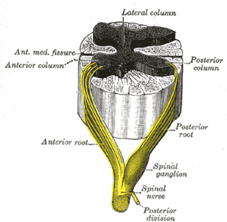
Dorsal root (sensory; its cell bodies are in the dorsal root ganglion, in the intervertebral foramen) and ventral root (motor; cell bodies in the ventral horn) unite in the foramen to form the short spinal nerve.
- Course
Exit rule: C1–C7 exit above the vertebra of the same number; C8 exits between C7 and T1; from T1 down each nerve exits below its same-numbered vertebra. Lower roots descend progressively farther within the canal before exiting (forming the cauda equina).
- Branches
- Dorsal (posterior) ramus: deep back muscles (e.g. Erector spinae), facet joints, and a strip of skin of the back
- Ventral (anterior) ramus: limbs and the anterolateral trunk; forms the cervical, brachial, lumbar and sacral plexuses, and the intercostal nerves
- Rami communicantes: white (preganglionic sympathetic, T1–L2 only) and gray (postganglionic, all levels) connect to the sympathetic trunk
- Meningeal (sinuvertebral) branch: back into the canal (dura, PLL, outer anulus)
- Motor
Via ventral roots (myotomes).
- Sensory
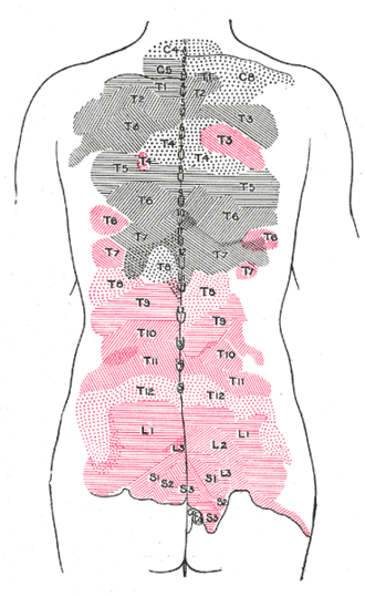
Via dorsal roots (dermatomes; e.g. C6 thumb, C7 middle finger, C8 little finger, T4 nipple, T10 umbilicus, L1 inguinal region, L4 medial leg, L5 dorsum of foot, S1 lateral foot).
- Notes
Lower limb root summary. L3: hip flexion/knee extension, anterior thigh. L4: knee extension, ankle dorsiflexion (tibialis anterior), patellar reflex, medial leg. L5: great-toe extension (EHL), hip abduction, dorsum of foot and first web space, no reliable common reflex. S1: plantarflexion, foot eversion, Achilles reflex, lateral foot and sole.
Muscles innervated
- Erector spinae — Dorsal (posterior) rami of the spinal nerves, segmentally.
Generated automatically from links in other entries.
Swipe sideways to see the whole diagram →


Clinical correlations
Radiculopathy PANCE
- Mechanism / lesion
Root compression or inflammation by disc herniation, foraminal stenosis (osteophytes, facet hypertrophy), or rarely tumor or infection.
- Classic presentation
Radiating pain, dermatomal sensory loss, myotomal weakness, and reduced reflexes in a single root distribution. Distinguished from peripheral nerve lesions by the pattern of affected muscles and skin.
Herpes zoster PANCE
- Mechanism / lesion
Reactivation of varicella-zoster virus latent in the dorsal root ganglion (or cranial nerve ganglia).
- Classic presentation
Prodromal pain then a unilateral dermatomal vesicular rash that doesn't cross the midline; possible postherpetic neuralgia.
High-yield PANCE pearls
- Root exit: C1–C7 above, C8 between C7–T1, T1 and below exit below their vertebra.
- Dermatomes: C6 thumb, C7 middle finger, C8 little finger, T4 nipple, T10 umbilicus, L4 medial leg, L5 dorsum of foot, S1 lateral foot.
- Reflexes: biceps C5–C6, brachioradialis C6, triceps C7, patellar L4, Achilles S1.
- Zoster lives in the dorsal root ganglion.
Imaging anatomy
- CT
CT (or CT myelography) for bony foraminal stenosis.
- MRI
MRI shows root compression in the foramen or lateral recess.
- Key views & landmarks
- EMG/nerve conduction studies: distinguish radiculopathy from plexopathy or peripheral neuropathy (sensory nerve action potentials are preserved in radiculopathy because the lesion is proximal to the dorsal root ganglion)
Physical exam
- Strength / function testing
Map motor weakness by myotome, sensory loss by dermatome, and reflexes by root; compare sides. Nerve tension tests (straight leg raise, Spurling) provoke radicular pain.
Self-quiz
Which spinal nerve exits through the intervertebral foramen between C7 and T1?
Correct answer: B
There are 8 cervical nerves but 7 cervical vertebrae, so C8 exits between C7 and T1. Below that, each nerve exits below its own vertebra.
Show answer
B
There are 8 cervical nerves but 7 cervical vertebrae, so C8 exits between C7 and T1. Below that, each nerve exits below its own vertebra.
Where are the cell bodies of sensory neurons carried in the spinal nerves located?
Correct answer: B
Sensory (pseudounipolar) neuron cell bodies lie in the dorsal root ganglion, the site where varicella-zoster virus lies latent.
Show answer
B
Sensory (pseudounipolar) neuron cell bodies lie in the dorsal root ganglion, the site where varicella-zoster virus lies latent.
Which reflex is most associated with the L4 root?
Correct answer: B
The patellar reflex tests L4 (with L3). The Achilles reflex tests S1.
Show answer
B
The patellar reflex tests L4 (with L3). The Achilles reflex tests S1.
White rami communicantes are found at which spinal levels?
Correct answer: C
White rami carry preganglionic sympathetic fibers and exist only at T1–L2, where the lateral horn is present. Gray rami (postganglionic) join every spinal nerve.
Show answer
C
White rami carry preganglionic sympathetic fibers and exist only at T1–L2, where the lateral horn is present. Gray rami (postganglionic) join every spinal nerve.
What does the dorsal (posterior) ramus of a typical spinal nerve supply?
Correct answer: E
The dorsal ramus supplies the intrinsic back muscles (e.g. erector spinae), the facet joints and a strip of skin of the back. The ventral ramus supplies the limbs and anterolateral trunk and forms the plexuses.
Show answer
E
The dorsal ramus supplies the intrinsic back muscles (e.g. erector spinae), the facet joints and a strip of skin of the back. The ventral ramus supplies the limbs and anterolateral trunk and forms the plexuses.