Axilla
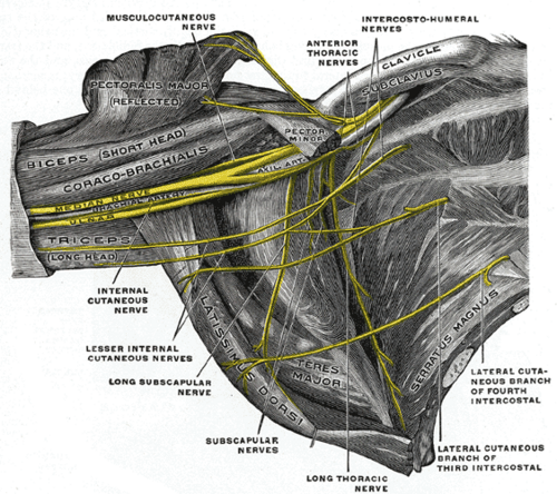
Pyramidal space between the arm and the thoracic wall. It is the passageway for the neurovascular bundle of the upper limb and contains the axillary lymph nodes, which drain most of the breast.
Core anatomy
- Location
Inferior to the glenohumeral joint, between the upper arm and the lateral thoracic wall.
- Boundaries
- apex
- Cervicoaxillary canal: clavicle (anterior), first rib (medial), superior border of the scapula (posterior)
- base
- Axillary fascia and skin of the armpit
- anterior wall
- Pectoralis major and pectoralis minor (with subclavius and the clavipectoral fascia)
- posterior wall
- Subscapularis (mainly), teres major and latissimus dorsi
- medial wall
- Thoracic wall (ribs 1–4 and intercostal muscles) covered by serratus anterior
- lateral wall
- Intertubercular groove of the humerus
- Contents
- Axillary artery and its branches
- Axillary vein and its tributaries
- Cords and branches of the brachial plexus
- Axillary lymph nodes (five groups: pectoral/anterior, subscapular/posterior, humeral/lateral, central, apical)
- Long thoracic nerve on the medial wall; thoracodorsal nerve on the posterior wall; intercostobrachial nerve crossing the space
- Axillary fat and the axillary sheath (surrounding the artery and plexus)
- Communications
With the posterior triangle of the neck through the apex (cervicoaxillary canal); with the arm; and with the scapular region through the quadrangular space, triangular space and triangular interval.
- Notes
Surgical levels of axillary nodes (by their relation to pectoralis minor): Level I lateral/inferior, Level II deep (posterior), Level III medial/superior (apical). Most lymph from the breast (about 75%) drains to the axillary nodes, first to the pectoral (anterior) group.
Mentioned in
- Axillary artery
- Axillary nerve
- Brachial plexus
- Breast
- Latissimus dorsi
- Long thoracic nerve
- Musculocutaneous nerve
- Pectoralis major
- Radial nerve
- Serratus anterior
- Subscapularis
- Triangles of the neck
Generated automatically from links in other entries.
Swipe sideways to see the whole diagram →

Clinical correlations
Axillary lymphadenopathy PANCE
- Mechanism / lesion
Drainage of the upper limb, breast and lateral trunk. Causes include infection (hand or arm cellulitis, cat-scratch disease), breast cancer metastasis, lymphoma and other malignancy.
- Classic presentation
Palpable nodes. Hard, fixed or matted nodes raise concern for malignancy. In breast cancer, axillary status determines staging; sentinel lymph node biopsy identifies the first node(s) draining the tumor.
Complications of axillary lymph node dissection PANCE
- Mechanism / lesion
Surgical injury to nerves on the walls of the axilla and disruption of lymphatic drainage.
- Classic presentation
Long thoracic nerve → winged scapula. Thoracodorsal nerve → weak latissimus dorsi (extension, adduction, medial rotation). Intercostobrachial nerve → numbness of the medial arm and axilla. Lymphedema of the upper limb.
Hidradenitis suppurativa
- Mechanism / lesion
Chronic inflammatory disease of follicles in intertriginous skin (axillae, groin).
- Classic presentation
Recurrent painful nodules, abscesses, sinus tracts and scarring in the axilla.
High-yield PANCE pearls
- Most breast lymph → axillary nodes (pectoral group first); level I–III defined by pectoralis minor.
- Axillary dissection injuries: long thoracic (winging), thoracodorsal (latissimus), intercostobrachial (medial arm numbness); lymphedema.
- Walls: anterior (pectorals), posterior (subscapularis, teres major, latissimus), medial (serratus anterior), lateral (intertubercular groove).
Imaging anatomy
- X-ray
Soft tissue only. Calcified nodes may be seen.
- CT
Contrast CT of the chest shows axillary adenopathy (size, necrosis) for lymphoma or cancer staging and axillary vascular injury.
- MRI
Breast MRI includes the axilla. MR neurography for the plexus.
- Ultrasound
First-line for axillary nodes. Normal nodes are oval with a thin cortex and a fatty echogenic hilum. Suspicious features are cortical thickening, loss of the fatty hilum, a round shape, and non-hilar blood flow. It guides core biopsy. The axillary artery is the landmark for axillary brachial plexus block.
- Key views & landmarks
- Mammography MLO view includes the low axilla
- Axillary ultrasound ± core biopsy
Physical exam
- Surface landmarks
Anterior axillary fold (lower border of pectoralis major) and posterior axillary fold (latissimus dorsi and teres major). The humeral head and axillary artery pulse can be felt in the lateral axilla.
- Palpation
With the patient's arm relaxed and supported by the examiner, reach high into the apex with cupped fingers and press against the chest wall. Then examine each group: central (against the chest wall), pectoral (inside the anterior fold), subscapular (inside the posterior fold), and lateral (along the upper humerus). Also check the infraclavicular and supraclavicular nodes.
- Strength / function testing
After axillary surgery, test serratus anterior (wall push), latissimus dorsi (resisted extension/adduction) and medial arm sensation.
Self-quiz
Two months after a left modified radical mastectomy with axillary dissection, a patient has numbness over the medial upper arm and axilla. Which nerve was most likely injured?
Correct answer: C
The intercostobrachial nerve (lateral cutaneous branch of T2) crosses the axilla to supply the skin of the medial arm and axilla. It is often injured in axillary dissection. The long thoracic and thoracodorsal nerves are motor nerves.
Show answer
C
The intercostobrachial nerve (lateral cutaneous branch of T2) crosses the axilla to supply the skin of the medial arm and axilla. It is often injured in axillary dissection. The long thoracic and thoracodorsal nerves are motor nerves.
Axillary lymph node levels are defined by their relationship to which muscle?
Correct answer: B
Level I: lateral/inferior to pectoralis minor. Level II: deep to it. Level III: medial/superior to it (apical).
Show answer
B
Level I: lateral/inferior to pectoralis minor. Level II: deep to it. Level III: medial/superior to it (apical).
Which muscle forms most of the posterior wall of the axilla?
Correct answer: C
The posterior wall is formed mainly by subscapularis, with teres major and latissimus dorsi inferiorly. Serratus anterior forms the medial wall.
Show answer
C
The posterior wall is formed mainly by subscapularis, with teres major and latissimus dorsi inferiorly. Serratus anterior forms the medial wall.
On ultrasound, which feature of an axillary lymph node is most suggestive of malignancy?
Correct answer: C
Cortical thickening and loss of the fatty hilum (plus a round shape and non-hilar vascularity) suggest metastatic involvement. The other options describe normal-appearing nodes.
Show answer
C
Cortical thickening and loss of the fatty hilum (plus a round shape and non-hilar vascularity) suggest metastatic involvement. The other options describe normal-appearing nodes.
After an axillary lymph node dissection, a patient's scapula lifts off the chest wall when she pushes against a wall. The injured nerve runs along which wall of the axilla?
Correct answer: C
The long thoracic nerve runs down the medial wall of the axilla on the surface of serratus anterior, so it is exposed during axillary dissection; injury causes medial winging. The thoracodorsal nerve runs on the posterior wall (latissimus dorsi), and the intercostobrachial nerve crosses the space (medial arm numbness).
Show answer
C
The long thoracic nerve runs down the medial wall of the axilla on the surface of serratus anterior, so it is exposed during axillary dissection; injury causes medial winging. The thoracodorsal nerve runs on the posterior wall (latissimus dorsi), and the intercostobrachial nerve crosses the space (medial arm numbness).