Urethra
Tube from the bladder to the exterior — about 18–20 cm and S-shaped in males, about 4 cm in females. The site of the injury determines where extravasated urine goes, and urethral injury must be excluded before catheterizing a trauma patient.
Core anatomy
- Location
From the internal urethral orifice at the bladder neck to the external urethral orifice (glans of the penis, or the vestibule between the clitoris and vaginal orifice).
- Parts
- Male — preprostatic (intramural) part: in the bladder neck, surrounded by the internal urethral sphincter
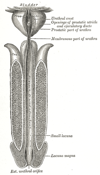
- Male — prostatic urethra: the widest and most dilatable part; urethral crest with the seminal colliculus, where the ejaculatory ducts open, and prostatic ducts in the sinuses on either side
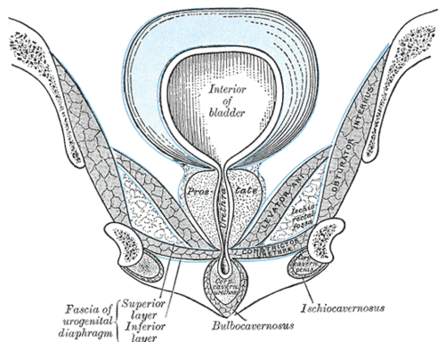
- Male — intermediate (membranous) urethra: the shortest and least distensible part, passing through the deep perineal pouch surrounded by the external urethral sphincter; the usual site of injury with pelvic fractures
- Male — spongy (penile) urethra: the longest part, in the corpus spongiosum; the ducts of the bulbourethral glands open into its bulbar portion; dilates in the glans as the navicular fossa
- Female urethra: about 4 cm, runs anteroinferiorly behind the symphysis, embedded in the anterior vaginal wall; paraurethral (Skene) glands open near its external orifice
- Relations
Male: passes through the prostate, the deep perineal pouch and the corpus spongiosum of the penis. Female: closely related to the anterior wall of the vagina.
- Arterial supply
Male: prostatic branches of the inferior vesical and middle rectal arteries (prostatic part); branches of the internal pudendal artery — artery of the bulb and urethral artery (membranous and spongy parts). Female: internal pudendal and vaginal arteries.
- Venous drainage
Prostatic or vesical venous plexuses and the internal pudendal veins.
- Lymphatic drainage
Proximal parts → internal iliac nodes; spongy urethra → mainly deep inguinal nodes (see pelvic and inguinal lymph drainage).
- Innervation
Prostatic/vesical plexuses (autonomic); somatic sensation and the external sphincter via the pudendal nerve (S2–S4).
- Notes
Catheterizing a male: hold the penis upward (perpendicular to the body) to straighten the prepubic curve; resistance is typical at the external sphincter (membranous part) and at an enlarged prostate. The external urethral orifice is the narrowest point.
Found in
Mentioned in
Generated automatically from links in other entries.
Swipe sideways to see the whole diagram →


Clinical correlations
Posterior (membranous) urethral injury PANCE
- Mechanism / lesion
Pelvic fracture (shearing at the prostatomembranous junction), usually in males.
- Classic presentation
Blood at the urethral meatus, inability to void, perineal or scrotal hematoma, and possibly a high-riding or non-palpable prostate on DRE (an unreliable sign). Urine extravasates into the deep perineal pouch and retropubic space.
- Notes
Do not pass a Foley catheter until a retrograde urethrogram excludes injury; a suprapubic catheter is used if the urethra is disrupted.
Anterior (bulbar) urethral injury PANCE
- Mechanism / lesion
Straddle injury (fall astride a bicycle bar or beam) crushing the bulbar urethra against the pubic arch.
- Classic presentation
Blood at the meatus and "butterfly" perineal bruising. If Buck fascia is torn, urine and blood spread in the superficial perineal pouch into the scrotum, penis and lower anterior abdominal wall — but not the thighs or anal triangle (see perineal pouches).
Urethritis PANCE
- Mechanism / lesion
Sexually transmitted infection — Neisseria gonorrhoeae, Chlamydia trachomatis, Mycoplasma genitalium, Trichomonas.
- Classic presentation
Dysuria and urethral discharge (purulent with gonorrhea). Diagnose with NAAT of first-catch urine. Treat gonorrhea with ceftriaxone 500 mg IM (plus doxycycline if chlamydia is not excluded) per CDC 2021 guidance; treat partners.
Hypospadias PANCE
- Mechanism / lesion
Incomplete fusion of the urethral folds, so the meatus opens on the ventral penis, scrotum or perineum. (Epispadias, a dorsal opening, is associated with bladder exstrophy.)
- Classic presentation
Abnormal meatus, dorsal hooded foreskin, often ventral curvature (chordee). Circumcision is avoided because the foreskin is used in the repair.
Posterior urethral valves
- Mechanism / lesion
Congenital obstructing membrane in the prostatic urethra of male infants.
- Classic presentation
The most common cause of bladder outlet obstruction in male newborns — bilateral hydronephrosis and oligohydramnios on prenatal ultrasound, poor stream. Diagnosed by VCUG.
High-yield PANCE pearls
- Pelvic fracture + blood at the meatus → retrograde urethrogram before Foley.
- Straddle injury → bulbar (anterior) urethra → extravasation into scrotum, penis, abdominal wall, not thighs.
- Hypospadias = ventral meatus (urethral folds) → no circumcision.
- Posterior urethral valves: male newborn, bilateral hydronephrosis, VCUG.
- Short female urethra → higher UTI risk.
Imaging anatomy
- X-ray
Retrograde urethrogram (RUG): contrast injected into the meatus under fluoroscopy shows extravasation (urethral injury) or stricture. VCUG: contrast instilled into the bladder, films during voiding — shows the posterior urethra (valves) and vesicoureteral reflux.
- MRI
Used for female urethral diverticulum and urethral tumors.
- Ultrasound
Sonourethrography can estimate the length of anterior urethral strictures.
- Key views & landmarks
- Retrograde urethrogram (oblique position)
- Voiding cystourethrogram
Physical exam
- Surface landmarks
The male bulbar urethra lies in the midline of the perineum, deep to bulbospongiosus; the female external urethral orifice lies between the clitoris and vaginal orifice.
- Palpation
Female — urethral tenderness or a suburethral mass expressing pus (diverticulum or Skene gland abscess).
- Strength / function testing
Inspect the meatus for blood, discharge, and position (hypospadias). In trauma check for perineal and scrotal hematoma and perform a DRE.
Self-quiz
A 30-year-old man is brought in after being crushed in a motor vehicle collision. He has a pelvic fracture, blood at the urethral meatus and a perineal hematoma. What is the most appropriate next step for his urinary tract?
Correct answer: B
Blood at the meatus with a pelvic fracture suggests posterior urethral injury. A retrograde urethrogram must come before any attempt at urethral catheterization, which could convert a partial into a complete tear.
Show answer
B
Blood at the meatus with a pelvic fracture suggests posterior urethral injury. A retrograde urethrogram must come before any attempt at urethral catheterization, which could convert a partial into a complete tear.
A 12-year-old boy falls astride the top bar of his bicycle. He develops swelling and bruising of the scrotum, penis and lower abdominal wall, but his thighs are spared. Which structure prevents spread into the thighs?
Correct answer: A
Urine from a bulbar urethral tear fills the superficial perineal pouch beneath Colles fascia, which is continuous with Scarpa fascia and dartos. The membranous layer fuses with the fascia lata just below the inguinal ligament, so fluid cannot enter the thigh; its fusion with the perineal membrane blocks spread into the anal triangle.
Show answer
A
Urine from a bulbar urethral tear fills the superficial perineal pouch beneath Colles fascia, which is continuous with Scarpa fascia and dartos. The membranous layer fuses with the fascia lata just below the inguinal ligament, so fluid cannot enter the thigh; its fusion with the perineal membrane blocks spread into the anal triangle.
Which is the shortest and least distensible part of the male urethra?
Correct answer: C
The intermediate (membranous) urethra (about 1–1.5 cm), surrounded by the external urethral sphincter in the deep perineal pouch, is the shortest and least distensible part (apart from the external orifice).
Show answer
C
The intermediate (membranous) urethra (about 1–1.5 cm), surrounded by the external urethral sphincter in the deep perineal pouch, is the shortest and least distensible part (apart from the external orifice).
A newborn boy's urethral meatus opens on the ventral surface of the penile shaft, and he has a dorsal hooded foreskin. What should the parents be advised?
Correct answer: C
This is hypospadias (incomplete fusion of the urethral folds, ventral meatus), often with a dorsal hooded foreskin and chordee. Circumcision is avoided because the foreskin is used in the repair. Epispadias is a dorsal opening associated with bladder exstrophy.
Show answer
C
This is hypospadias (incomplete fusion of the urethral folds, ventral meatus), often with a dorsal hooded foreskin and chordee. Circumcision is avoided because the foreskin is used in the repair. Epispadias is a dorsal opening associated with bladder exstrophy.
A male newborn had bilateral hydronephrosis and oligohydramnios on prenatal ultrasound and now has a weak urinary stream. Which study confirms the most likely diagnosis?
Correct answer: E
Posterior urethral valves (an obstructing membrane in the prostatic urethra) are the most common cause of bladder outlet obstruction in male newborns and are diagnosed by VCUG.
Show answer
E
Posterior urethral valves (an obstructing membrane in the prostatic urethra) are the most common cause of bladder outlet obstruction in male newborns and are diagnosed by VCUG.