Internal iliac artery
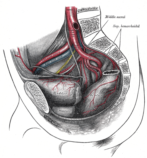
Main artery of the pelvis, supplying the pelvic viscera, perineum, gluteal region and medial thigh. Its branches bleed in pelvic fractures and are embolized for postpartum hemorrhage and fibroids.
Core anatomy
- Location
Lateral pelvic wall, from the pelvic brim to the upper border of the greater sciatic foramen.
- Origin
Bifurcation of the common iliac artery at about the level of the L5/S1 disc, anterior to the sacroiliac joint.
- Course
Descends posteromedially into the lesser pelvis, with the ureter anterior to it and the internal iliac vein and lumbosacral trunk posterior, and divides near the upper margin of the greater sciatic foramen into anterior and posterior divisions (the branching pattern varies considerably).
- Branches
- Posterior division (mnemonic "I Love Sex"): iliolumbar, lateral sacral, superior gluteal (largest branch; leaves through the greater sciatic foramen above piriformis)
- Anterior division — visceral: umbilical (patent part gives the superior vesical arteries; its obliterated part is the medial umbilical ligament); inferior vesical (male) or vaginal (female); uterine artery (female); middle rectal
- Anterior division — parietal: obturator (through the obturator canal to the medial thigh); internal pudendal (out through the greater and back in through the lesser sciatic foramen, into the pudendal canal to supply the perineum, anal canal and erectile tissue); inferior gluteal (below piriformis)
- Supplies
Pelvic viscera (urinary bladder, rectum, prostate, uterus, vagina), the perineum and external genitalia, the gluteal muscles, the medial thigh (obturator) and the pelvic walls.
- Anastomoses
Iliolumbar ↔ lumbar arteries; lateral sacral ↔ median sacral; middle rectal ↔ superior rectal (from the inferior mesenteric artery) and inferior rectal; uterine ↔ ovarian; obturator ↔ inferior epigastric (pubic branches); inferior gluteal ↔ medial and lateral circumflex femoral (cruciate anastomosis). These collaterals allow bilateral internal iliac ligation or embolization without pelvic necrosis.
- Termination
Divides into its anterior and posterior divisions.
- Relations
An accessory (aberrant) obturator artery from the inferior epigastric or external iliac artery crosses the superior pubic ramus in a sizable minority of people ("corona mortis") and can bleed during hernia repair or pelvic fracture fixation. verify
Branches
Structures supplied
- Ductus deferens and seminal glands
- Gluteus maximus
- Gluteus medius and minimus
- Pelvic diaphragm
- Penis
- Prostate
- Rectum
- Sacroiliac joint
- Urinary bladder
- Uterus
- Vagina
Mentioned in
Generated automatically from links in other entries.
Swipe sideways to see the whole diagram →

Clinical correlations
Arterial hemorrhage in pelvic fractures PANCE
- Mechanism / lesion
Shearing of branches by fracture fragments — the superior gluteal (posterior ring and sacroiliac injuries), internal pudendal and obturator arteries are most often involved.
- Classic presentation
Ongoing hemodynamic instability despite a binder and resuscitation; an active contrast blush on CT. Treated with angioembolization (or preperitoneal packing). See bony pelvis.
Postpartum hemorrhage — embolization or ligation
- Mechanism / lesion
Uterine atony or trauma not controlled by uterotonics.
- Classic presentation
Uterine artery embolization, or ligation of the uterine or internal iliac arteries, reduces pulse pressure to the uterus while collaterals preserve perfusion.
Aortoiliac occlusive disease (Leriche syndrome) PANCE
- Mechanism / lesion
Atherosclerotic occlusion of the distal aorta and/or both iliac arteries.
- Classic presentation
Buttock and thigh claudication, erectile dysfunction (reduced internal pudendal flow), and diminished or absent femoral pulses.
High-yield PANCE pearls
- Leriche syndrome triad: buttock claudication, impotence, absent femoral pulses.
- Superior gluteal artery = largest branch; exits above piriformis; injured in posterior pelvic fractures.
- Posterior division: Iliolumbar, Lateral sacral, Superior gluteal.
- Obliterated umbilical artery = medial umbilical ligament.
Imaging anatomy
- X-ray
Conventional catheter angiography is used for diagnosis and embolization (trauma, PPH, uterine fibroids, tumor bleeding).
- CT
CT angiography shows active extravasation in trauma, aneurysms and occlusive disease. Internal iliac aneurysms are often associated with abdominal aortic aneurysms.
- Ultrasound
Duplex ultrasound of the iliac arteries assesses aortoiliac disease (with ankle-brachial indices).
- Key views & landmarks
- CT angiography of the abdomen and pelvis
- Pelvic angiography with selective internal iliac injection
Physical exam
- Surface landmarks
Not palpable. Iliac inflow is judged from the femoral pulses (just below the midpoint of the inguinal ligament).
- Strength / function testing
Compare femoral pulses, listen for iliac/femoral bruits, and measure the ankle-brachial index; ask about buttock claudication and erectile dysfunction.
Self-quiz
A 62-year-old man who smokes has cramping pain in both buttocks and thighs when walking two blocks, relieved by rest. He also reports erectile dysfunction. Femoral pulses are weak bilaterally. Where is the most likely site of arterial occlusion?
Correct answer: C
Leriche syndrome (aortoiliac occlusive disease) causes buttock and thigh claudication, erectile dysfunction (reduced flow to the internal iliac/internal pudendal arteries) and diminished femoral pulses. Superficial femoral disease causes calf claudication.
Show answer
C
Leriche syndrome (aortoiliac occlusive disease) causes buttock and thigh claudication, erectile dysfunction (reduced flow to the internal iliac/internal pudendal arteries) and diminished femoral pulses. Superficial femoral disease causes calf claudication.
Which artery leaves the pelvis through the greater sciatic foramen above the piriformis muscle?
Correct answer: C
The superior gluteal artery (with the superior gluteal nerve) is the only structure passing above piriformis. The inferior gluteal and internal pudendal arteries pass below it.
Show answer
C
The superior gluteal artery (with the superior gluteal nerve) is the only structure passing above piriformis. The inferior gluteal and internal pudendal arteries pass below it.
The medial umbilical ligament is a remnant of which fetal vessel?
Correct answer: B
The distal part of each umbilical artery becomes a medial umbilical ligament; the proximal part stays patent and gives the superior vesical arteries. The urachus becomes the median umbilical ligament, and the umbilical vein the ligamentum teres.
Show answer
B
The distal part of each umbilical artery becomes a medial umbilical ligament; the proximal part stays patent and gives the superior vesical arteries. The urachus becomes the median umbilical ligament, and the umbilical vein the ligamentum teres.
Which artery is a branch of the posterior division of the internal iliac artery?
Correct answer: E
The posterior division gives the iliolumbar, lateral sacral and superior gluteal arteries (mnemonic "I Love Sex"). The obturator, internal pudendal, inferior gluteal and uterine arteries come from the anterior division.
Show answer
E
The posterior division gives the iliolumbar, lateral sacral and superior gluteal arteries (mnemonic "I Love Sex"). The obturator, internal pudendal, inferior gluteal and uterine arteries come from the anterior division.
Which artery leaves the pelvis through the greater sciatic foramen, re-enters through the lesser sciatic foramen, and runs in the pudendal canal to supply the perineum and erectile tissue?
Correct answer: A
The internal pudendal artery (anterior division) exits through the greater sciatic foramen, re-enters through the lesser sciatic foramen, and runs in the pudendal (Alcock) canal to supply the perineum, the anal canal below the pectinate line and the erectile tissue. Reduced flow in it contributes to erectile dysfunction in Leriche syndrome.
Show answer
A
The internal pudendal artery (anterior division) exits through the greater sciatic foramen, re-enters through the lesser sciatic foramen, and runs in the pudendal (Alcock) canal to supply the perineum, the anal canal below the pectinate line and the erectile tissue. Reduced flow in it contributes to erectile dysfunction in Leriche syndrome.