Heart
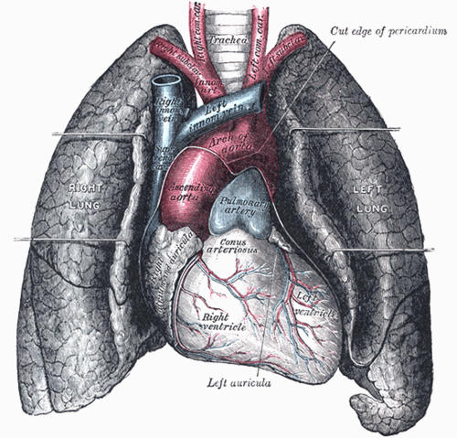
Four-chambered muscular pump in the middle mediastinum. Knowing which chamber forms each surface and border is the key to exam findings, ECGs and chest X-rays.
Core anatomy
- Location
Middle mediastinum, within the pericardium, lying obliquely with about two-thirds to the left of the midline. The apex points anteriorly, inferiorly and to the left, at about the left 5th intercostal space in the midclavicular line.
- Parts
- Right atrium: receives the superior vena cava, inferior vena cava and coronary sinus. Contains the SA node (near the SVC opening), the fossa ovalis, and the AV node (in the interatrial septum near the coronary sinus)
- Right ventricle: trabeculae carneae, septomarginal trabecula (moderator band carrying the right bundle branch), tricuspid valve inflow, pulmonary valve outflow via the conus arteriosus (infundibulum)
- Left atrium: receives four pulmonary veins. The most posterior chamber, directly in front of the esophagus
- Left ventricle: thickest wall. Mitral valve inflow, aortic valve outflow. Forms the apex
- Interatrial and interventricular septa (the upper interventricular septum is membranous)
- Conduction system: see cardiac conduction system
- Relations
Surfaces. Anterior (sternocostal): mainly the right ventricle. Diaphragmatic (inferior): mainly the left ventricle, with part of the right ventricle. Base (posterior): mainly the left atrium. Borders on the PA chest X-ray. Right border: SVC (above) and right atrium. Left border, from top to bottom: aortic knob (arch), pulmonary trunk, left atrial appendage (auricle), left ventricle. The right ventricle does not form a border on a PA film.
- Arterial supply
Right coronary artery and left coronary artery (via the anterior interventricular artery and circumflex artery). The coronary arteries fill mainly during diastole.
- Venous drainage
Cardiac veins → coronary sinus (in the posterior atrioventricular groove) → right atrium. Small anterior cardiac veins drain directly into the right atrium.
- Innervation
Cardiac plexus. Sympathetic (upper thoracic segments, about T1–T4/T5): increases heart rate and contractility; carries visceral pain, which is referred to the chest, left arm and jaw. Parasympathetic (vagus): slows the heart rate.
Found in
Mentioned in
Generated automatically from links in other entries.

Clinical correlations
Penetrating cardiac injury PANCE
- Mechanism / lesion
The right ventricle forms most of the anterior surface, so it's the chamber most often injured by anterior stab wounds.
- Classic presentation
Hemopericardium with cardiac tamponade (see pericardium) or hemorrhagic shock.
Left atrial enlargement PANCE
- Mechanism / lesion
Mitral stenosis or regurgitation, or long-standing atrial fibrillation.
- Classic presentation
The enlarged posterior chamber can compress the esophagus (dysphagia) or, rarely, the left recurrent laryngeal nerve (hoarseness, Ortner syndrome). Predisposes to atrial fibrillation and thrombus in the left atrial appendage.
Referred cardiac pain (angina / MI) PANCE
- Mechanism / lesion
Visceral afferents travel with sympathetic fibers to upper thoracic spinal segments, which also receive somatic input from the chest wall and medial arm.
- Classic presentation
Pressure-like retrosternal pain radiating to the left arm (T1–T2 dermatomes), jaw or epigastrium. Atypical presentations are more common in women, older adults and people with diabetes.
High-yield PANCE pearls
- Apex / PMI: left 5th intercostal space, midclavicular line. A laterally displaced PMI suggests LV dilation.
- Anterior surface = right ventricle; posterior (base) = left atrium.
- PA CXR left border (top → bottom): aortic knob, pulmonary trunk, left atrial appendage, left ventricle.
- Coronary perfusion occurs mainly in diastole; tachycardia shortens diastole.
- Enlarged left atrium → dysphagia (esophagus) ± hoarseness (Ortner syndrome); TEE images the LA best because it sits right behind the esophagus.
Imaging anatomy
- X-ray
Cardiothoracic ratio on a PA film: greater than about 0.5 suggests cardiomegaly (AP films exaggerate heart size). LA enlargement: a double density behind the right heart, a straightened left heart border, and a widened carina. RV enlargement: filling of the retrosternal space on the lateral view.
- CT
Cardiac CT and coronary CT angiography show coronary anatomy and calcium; CT also shows pericardial disease.
- MRI
Cardiac MRI is the reference standard for ventricular volumes and function and for tissue characterization (late gadolinium enhancement in infarction and cardiomyopathy).
- Ultrasound
Echocardiography is first-line for chambers, walls, valves and function. Standard transthoracic windows: parasternal long and short axis, apical four-chamber, subcostal, suprasternal. TEE (transesophageal) gives better views of the posterior structures: left atrium and appendage (thrombus), mitral valve, aorta.
- Key views & landmarks
- PA and lateral CXR
- Transthoracic echo: parasternal long/short axis, apical 4-chamber, subcostal (FAST view for pericardial fluid)
Physical exam
- Surface landmarks
Surface projection: right border from the 3rd to the 6th right costal cartilage about 1–2 cm from the sternum; left border from the 2nd left intercostal space to the apex (5th intercostal space, midclavicular line). verify
- Palpation
Point of maximal impulse (PMI) at the apex: normally a small (about 1–2 cm) tap. Displacement or enlargement suggests LV dilation. A parasternal heave suggests RV enlargement. Thrills are palpable murmurs.
- Strength / function testing
Auscultate at the four classic areas (aortic, pulmonic, tricuspid, mitral) and Erb's point, with the diaphragm and the bell, supine and in the left lateral decubitus position (mitral), and sitting forward (aortic regurgitation). See the valve entries.
Self-quiz
A 25-year-old man has a stab wound just left of the sternum at the 4th intercostal space. Which cardiac chamber is most likely injured?
Correct answer: B
The right ventricle forms most of the sternocostal (anterior) surface of the heart, so it's the chamber most at risk from anterior penetrating wounds.
Show answer
B
The right ventricle forms most of the sternocostal (anterior) surface of the heart, so it's the chamber most at risk from anterior penetrating wounds.
A patient with long-standing mitral stenosis develops difficulty swallowing solids. Which chamber is compressing the esophagus?
Correct answer: C
The left atrium is the most posterior chamber, lying directly in front of the esophagus. Mitral stenosis enlarges it, which can indent the esophagus.
Show answer
C
The left atrium is the most posterior chamber, lying directly in front of the esophagus. Mitral stenosis enlarges it, which can indent the esophagus.
Which structure forms most of the right border of the heart on a PA chest radiograph?
Correct answer: B
The right atrium forms the right heart border, with the SVC above it. The right ventricle is anterior and doesn't form a border on the PA view.
Show answer
B
The right atrium forms the right heart border, with the SVC above it. The right ventricle is anterior and doesn't form a border on the PA view.
Where is the normal apical impulse (PMI) located in an adult?
Correct answer: D
The apex beat is normally at the left 5th intercostal space, midclavicular line. Displacement laterally or inferiorly suggests left ventricular enlargement.
Show answer
D
The apex beat is normally at the left 5th intercostal space, midclavicular line. Displacement laterally or inferiorly suggests left ventricular enlargement.
On a PA chest radiograph, which structures form the left heart border from top to bottom?
Correct answer: C
The left border, from top to bottom, is the aortic knob, pulmonary trunk, left atrial appendage and left ventricle. The right border is the SVC and right atrium. The right ventricle forms the anterior surface and doesn't form a border on a PA film.
Show answer
C
The left border, from top to bottom, is the aortic knob, pulmonary trunk, left atrial appendage and left ventricle. The right border is the SVC and right atrium. The right ventricle forms the anterior surface and doesn't form a border on a PA film.