Great saphenous vein
The longest vein in the body, running superficially from the front of the medial malleolus to the groin. It is the usual source of varicose veins and a common graft for coronary bypass.
Core anatomy
- Location
Superficial fascia of the medial leg and thigh.
- Formation
From the medial end of the dorsal venous arch of the foot.
- Course
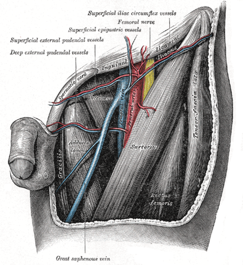
Passes anterior to the medial malleolus (reliably, for cutdown access), ascends along the medial leg with the saphenous nerve, passes a hand's breadth behind the medial border of the patella, up the medial thigh, and through the saphenous opening in the fascia lata to join the femoral vein about 3–4 cm below and lateral to the pubic tubercle (the saphenofemoral junction).
- Tributaries
- Superficial veins of the medial leg and thigh, including accessory saphenous veins
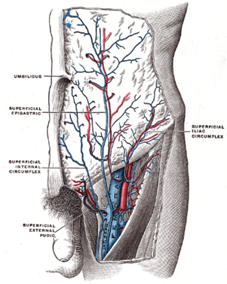
- Near the junction: superficial epigastric, superficial circumflex iliac and superficial external pudendal veins
- Communications with the small saphenous vein and, through perforating veins with one-way valves, with the deep veins
- Drains into
Femoral vein at the saphenofemoral junction (see deep veins of the lower limb).
- Relations
The saphenous nerve runs with it below the knee (injury causes medial leg numbness). The small saphenous vein runs up the back of the calf to the popliteal vein.
Found in
Mentioned in
Generated automatically from links in other entries.
Swipe sideways to see the whole diagram →


Clinical correlations
Varicose veins and chronic venous insufficiency PANCE
- Mechanism / lesion
Valve incompetence (often at the saphenofemoral junction) with reflux and venous hypertension; risk factors — age, female sex, pregnancy, obesity, prolonged standing, prior DVT.
- Classic presentation
Dilated tortuous veins, aching and heaviness worse at the end of the day, ankle edema; advanced disease causes hyperpigmentation, lipodermatosclerosis and venous ulcers above the medial malleolus. Treat with compression and leg elevation; endovenous ablation or sclerotherapy for symptomatic reflux.
Superficial thrombophlebitis PANCE
- Mechanism / lesion
Thrombosis of a superficial vein (varicosities, IV cannulas, pregnancy, malignancy).
- Classic presentation
A tender, red, palpable cord. Ultrasound because about a quarter have associated DVT; thrombus within about 3 cm of the saphenofemoral junction (or long segments) is treated with anticoagulation.
Saphenous vein graft
- Mechanism / lesion
Harvested for coronary artery bypass grafting and peripheral bypass (reversed so its valves don't block flow).
- Classic presentation
Donor-leg complications include wound infection and saphenous nerve numbness.
High-yield PANCE pearls
- Venous ulcer: above the medial malleolus, shallow, irregular, with hyperpigmentation → compression.
- Arterial ulcer: distal (toes, lateral malleolus), punched out, painful.
- Superficial thrombophlebitis → ultrasound (DVT coexists); anticoagulate if near the saphenofemoral junction.
- GSV lies anterior to the medial malleolus.
Imaging anatomy
- Ultrasound
Venous duplex ultrasound with reflux testing (standing) maps incompetent valves and excludes DVT.
- Key views & landmarks
- Venous reflux duplex ultrasound (standing)
Physical exam
- Surface landmarks
Visible or palpable just in front of the medial malleolus; the saphenofemoral junction lies about 3–4 cm inferolateral to the pubic tubercle.
- Palpation
Palpate for cords, tenderness and a cough impulse at the saphenofemoral junction (reflux).
- Strength / function testing
Inspect the standing patient for varicosities, edema, skin changes and ulcers; compare with arterial pulses before compression therapy (check ABI).
Trendelenburg (Brodie–Trendelenburg) test
- Technique
Elevate the leg to empty the veins, apply a tourniquet around the upper thigh (or press on the saphenofemoral junction), then have the patient stand.
- Positive test
Rapid filling of varicosities from above when the tourniquet is released (saphenofemoral incompetence); filling with the tourniquet in place implies incompetent perforators below.
- Significance
Localizes valvular incompetence (largely replaced by duplex ultrasound).
Self-quiz
A 62-year-old woman who works as a cashier has a shallow, irregular ulcer above her left medial malleolus with surrounding brown pigmentation and ankle edema. Pedal pulses are normal. What is the first-line treatment?
Correct answer: B
This is a venous stasis ulcer from chronic venous insufficiency. Compression (after confirming adequate arterial flow) and elevation are the cornerstone; ablation of refluxing veins reduces recurrence.
Show answer
B
This is a venous stasis ulcer from chronic venous insufficiency. Compression (after confirming adequate arterial flow) and elevation are the cornerstone; ablation of refluxing veins reduces recurrence.
Where does the great saphenous vein end?
Correct answer: B
The great saphenous vein passes through the saphenous opening in the fascia lata to join the femoral vein in the femoral triangle. The small saphenous vein drains to the popliteal vein.
Show answer
B
The great saphenous vein passes through the saphenous opening in the fascia lata to join the femoral vein in the femoral triangle. The small saphenous vein drains to the popliteal vein.
Where is the great saphenous vein reliably found at the ankle, for example for a venous cutdown?
Correct answer: C
The great saphenous vein begins at the medial end of the dorsal venous arch and passes anterior to the medial malleolus, a reliable site for cutdown. The small saphenous vein passes behind the lateral malleolus.
Show answer
C
The great saphenous vein begins at the medial end of the dorsal venous arch and passes anterior to the medial malleolus, a reliable site for cutdown. The small saphenous vein passes behind the lateral malleolus.
Which feature suggests an arterial rather than a venous leg ulcer?
Correct answer: D
Arterial ulcers are distal (toes, lateral malleolus), punched out and painful, with poor pulses. Venous ulcers are shallow and irregular, above the medial malleolus, with hyperpigmentation and edema, and are treated with compression.
Show answer
D
Arterial ulcers are distal (toes, lateral malleolus), punched out and painful, with poor pulses. Venous ulcers are shallow and irregular, above the medial malleolus, with hyperpigmentation and edema, and are treated with compression.
A patient has a tender, red, palpable cord along the great saphenous vein in the thigh. What is the most appropriate next step?
Correct answer: B
Superficial thrombophlebitis can coexist with DVT (in about a quarter of cases), so duplex ultrasound is needed. Thrombus close to the saphenofemoral junction or long segments are treated with anticoagulation.
Show answer
B
Superficial thrombophlebitis can coexist with DVT (in about a quarter of cases), so duplex ultrasound is needed. Thrombus close to the saphenofemoral junction or long segments are treated with anticoagulation.