Inferior vena cava
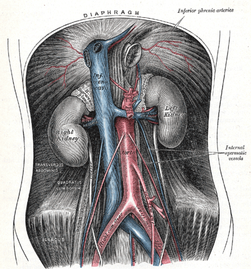
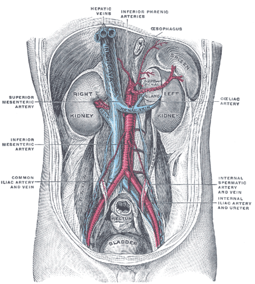
Largest vein of the body, returning blood from below the diaphragm to the right atrium. It lies to the right of the aorta, passes through the diaphragm at T8, and is compressed by the gravid uterus in the supine position.
Core anatomy
- Location
Retroperitoneal, to the right of the abdominal aorta on the lumbar vertebral bodies, then in a groove on the posterior surface of the liver.
- Formation
Union of the right and left common iliac veins at about L5, behind the right common iliac artery.
- Course
Ascends to the right of the aorta, passes through the caval opening of the central tendon of the diaphragm at T8, and enters the right atrium.
- Tributaries
- Common iliac veins
- Lumbar veins (3rd and 4th)
- Right gonadal vein (the left drains into the left renal vein)
- Renal veins: the left renal vein is longer, crosses anterior to the aorta just below the SMA, and receives the left gonadal and left suprarenal veins
- Right suprarenal vein (short, directly into the IVC)
- Inferior phrenic veins
- Hepatic veins (just below the diaphragm)
- Drains into
Right atrium of the heart.
- Relations
Anterior: right common iliac artery, root of the mesentery, 3rd part of the duodenum, head of the pancreas, the portal vein and the omental foramen (lesser sac), and the liver. Posterior: right sympathetic trunk, right renal artery, right crus of the diaphragm.
Tributaries
Mentioned in
Generated automatically from links in other entries.


Clinical correlations
Supine hypotensive syndrome of pregnancy (aortocaval compression) PANCE
- Mechanism / lesion
The gravid uterus compresses the IVC (and aorta) when the patient lies supine, typically after about 20 weeks, reducing venous return.
- Classic presentation
Hypotension, dizziness, pallor or fetal distress when supine, relieved by left lateral tilt. Manual left uterine displacement during maternal resuscitation.
IVC filter PANCE
- Mechanism / lesion
A filter placed in the infrarenal IVC traps emboli from the legs.
- Classic presentation
Indicated for acute proximal DVT or PE when anticoagulation is contraindicated (or has failed). Removable filters should be retrieved when no longer needed.
Tumor extension and obstruction
- Mechanism / lesion
Renal cell carcinoma can grow into the renal vein and IVC as a tumor thrombus; IVC thrombosis or compression.
- Classic presentation
Bilateral leg edema, dilated abdominal wall collateral veins (flow upward), a right-sided varicocele, and Budd–Chiari physiology if the thrombus reaches the hepatic segment.
High-yield PANCE pearls
- IVC forms at L5, passes the diaphragm at T8.
- Right gonadal vein → IVC; left gonadal vein → left renal vein.
- Pregnant patient hypotensive when supine → left lateral positioning (aortocaval compression).
- IVC filter when anticoagulation is contraindicated in DVT/PE.
Imaging anatomy
- CT
Contrast CT shows thrombus, tumor extension (RCC), congenital anomalies (duplicated or left-sided IVC, azygos continuation), and filter position.
- Ultrasound
Visible in the subcostal view. IVC diameter and inspiratory collapse are used at the bedside as a rough guide to right atrial pressure and fluid status (limited reliability in ventilated patients). Doppler for thrombosis. verify
- Key views & landmarks
- Subcostal long-axis view of the IVC entering the right atrium
- CT venography
Physical exam
- Surface landmarks
Lies to the right of the midline on the lumbar spine; not palpable.
- Strength / function testing
Look for bilateral leg edema and abdominal wall collateral veins. In IVC obstruction, blood in the periumbilical collaterals flows upward; in portal hypertension (caput medusae), it flows away from the umbilicus.
Self-quiz
A 32-year-old woman at 34 weeks' gestation becomes lightheaded and hypotensive while lying flat for an ultrasound. What is the first intervention?
Correct answer: B
Aortocaval compression by the gravid uterus reduces venous return. Left lateral tilt (or manual left uterine displacement) relieves it.
Show answer
B
Aortocaval compression by the gravid uterus reduces venous return. Left lateral tilt (or manual left uterine displacement) relieves it.
The left gonadal vein drains into which vessel?
Correct answer: B
The left gonadal vein drains into the left renal vein; the right gonadal vein drains directly into the IVC.
Show answer
B
The left gonadal vein drains into the left renal vein; the right gonadal vein drains directly into the IVC.
A patient with a proximal DVT has an intracranial hemorrhage. What is the most appropriate way to prevent pulmonary embolism?
Correct answer: C
When anticoagulation is contraindicated (active bleeding), an IVC filter can prevent PE from a leg DVT.
Show answer
C
When anticoagulation is contraindicated (active bleeding), an IVC filter can prevent PE from a leg DVT.
The inferior vena cava passes through the diaphragm at which opening and level?
Correct answer: B
The IVC passes through the caval opening in the central tendon at T8 ("I 8 10 Eggs At 12": IVC T8, esophagus T10, aorta T12).
Show answer
B
The IVC passes through the caval opening in the central tendon at T8 ("I 8 10 Eggs At 12": IVC T8, esophagus T10, aorta T12).
A 62-year-old man develops a new right-sided varicocele that does not decompress when he lies down. What should this prompt?
Correct answer: E
The right testicular vein drains directly into the IVC, so a new right-sided or non-decompressing varicocele suggests obstruction (e.g. renal cell carcinoma tumor thrombus in the IVC or a retroperitoneal mass). Most varicoceles are left-sided.
Show answer
E
The right testicular vein drains directly into the IVC, so a new right-sided or non-decompressing varicocele suggests obstruction (e.g. renal cell carcinoma tumor thrombus in the IVC or a retroperitoneal mass). Most varicoceles are left-sided.