Mediastinum
Central compartment of the thorax between the pleural cavities, divided into superior and inferior parts. Knowing what is in each compartment leads you to the differential for a mediastinal mass.
Core anatomy
- Location
Between the two pleural cavities, from the thoracic inlet above to the diaphragm below, and from the sternum in front to the thoracic vertebrae behind.
- Boundaries
- superior
- Superior thoracic aperture (thoracic inlet)
- inferior
- Diaphragm
- anterior
- Sternum and costal cartilages
- posterior
- Bodies of the thoracic vertebrae
- lateral
- Mediastinal parietal pleura on each side
- internal division
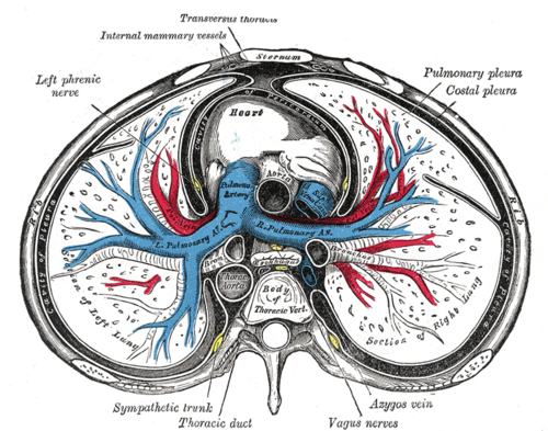
- The transverse thoracic plane through the sternal angle and the T4/T5 disc separates the superior mediastinum from the inferior mediastinum, which the pericardium divides into anterior, middle and posterior parts.
- Contents
- Superior: Thymus (or remnant), great veins (brachiocephalic veins, upper superior vena cava), aortic arch and its branches, trachea, esophagus, thoracic duct, vagus and phrenic nerves, left recurrent laryngeal nerve
- Anterior (between the sternum and pericardium): thymus remnants in children, fat, lymph nodes, sternopericardial ligaments, branches of the internal thoracic vessels
- Middle: Heart and pericardium, ascending aorta, pulmonary trunk (pulmonary arteries), lower SVC, main bronchi and the tracheal bifurcation, phrenic nerves
- Posterior (behind the pericardium): descending thoracic aorta, esophagus, thoracic duct, azygos vein and hemiazygos veins, vagus nerves, sympathetic trunks and splanchnic nerves, posterior mediastinal lymph nodes
- Communications
With the neck (through the thoracic inlet, so neck infections and air can spread into it) and the abdomen (through the diaphragmatic openings).
- Notes
Radiologists increasingly use the ITMIG three-compartment model: prevascular (roughly anterior), visceral (roughly middle), and paravertebral (roughly posterior), based on CT landmarks. The classic masses map similarly.
Mentioned in
Generated automatically from links in other entries.
Swipe sideways to see the whole diagram →

Clinical correlations
Mediastinal masses by compartment PANCE
- Mechanism / lesion
Anterior (4 T's): thymoma, teratoma/germ cell tumor, thyroid (retrosternal goiter), "terrible" lymphoma. Middle: lymphadenopathy (lymphoma, sarcoidosis, metastases, TB), bronchogenic and pericardial cysts, aortic aneurysm. Posterior: neurogenic tumors (schwannoma and neurofibroma in adults, neuroblastoma in children), esophageal lesions, descending aortic aneurysm.
- Classic presentation
Often incidental. Compression of adjacent structures causes cough, stridor, dysphagia, SVC syndrome, hoarseness (recurrent laryngeal nerve), or Horner syndrome.
Widened mediastinum PANCE
- Mechanism / lesion
Aortic dissection or traumatic aortic injury (mediastinal hematoma), lymphadenopathy (lymphoma, sarcoidosis, inhalational anthrax), masses.
- Classic presentation
Seen on CXR. In trauma or tearing chest pain it demands CT angiography.
Pneumomediastinum and mediastinitis
- Mechanism / lesion
Pneumomediastinum: alveolar rupture (asthma, Valsalva, drug inhalation), esophageal perforation, or airway injury. Mediastinitis: esophageal perforation, post-sternotomy infection, or descending spread of neck infection.
- Classic presentation
Chest pain, subcutaneous emphysema, Hamman crunch. Mediastinitis adds fever, sepsis and a widened mediastinum; it is life-threatening.
High-yield PANCE pearls
- Sternal angle plane (T4/T5) = superior/inferior mediastinum boundary.
- Anterior: 4 T's. Middle: lymph nodes, cysts. Posterior: neurogenic tumors.
- Widened mediastinum → aortic dissection/injury until proven otherwise; also lymphoma and anthrax.
- Hamman crunch = pneumomediastinum.
Imaging anatomy
- X-ray
PA and lateral CXR. The lateral film localizes masses (retrosternal = anterior; overlying the heart = middle; over the spine = posterior). The hilum overlay sign (hilar vessels visible through a mass) suggests an anterior or posterior mass rather than hilar enlargement. The cervicothoracic sign (a mass whose upper border is sharp above the clavicles) places it posteriorly.
- CT
Contrast CT is the main modality for localization, characterization (fat, calcium, cystic vs solid, vascular), and biopsy planning.
- MRI
MRI for posterior (neurogenic) masses with possible spinal canal extension, and for distinguishing cysts and thymic hyperplasia.
- Key views & landmarks
- PA and lateral CXR
- CT chest with IV contrast
Physical exam
- Surface landmarks
The sternal angle marks the boundary between the superior and inferior mediastinum.
- Strength / function testing
Look for signs of compression: facial swelling and neck vein distension (SVC), stridor (trachea), dysphagia (esophagus), hoarseness (left recurrent laryngeal nerve), ptosis and miosis (Horner syndrome, sympathetic chain). Palpate cervical and supraclavicular lymph nodes. Auscultate for Hamman crunch.
Self-quiz
A 30-year-old man has an incidental mass on CXR that overlies the vertebral bodies on the lateral view. CT shows a well-circumscribed paravertebral mass widening a neural foramen. What is the most likely diagnosis?
Correct answer: C
A posterior (paravertebral) mediastinal mass with foraminal widening is typically a neurogenic tumor such as a schwannoma or neurofibroma in adults.
Show answer
C
A posterior (paravertebral) mediastinal mass with foraminal widening is typically a neurogenic tumor such as a schwannoma or neurofibroma in adults.
The transverse thoracic plane separating the superior and inferior mediastinum passes through which vertebral level?
Correct answer: C
The plane through the sternal angle and the T4/T5 disc divides the superior mediastinum from the inferior.
Show answer
C
The plane through the sternal angle and the T4/T5 disc divides the superior mediastinum from the inferior.
Which structure is located in the middle mediastinum?
Correct answer: D
The middle mediastinum contains the heart and pericardium, the roots of the great vessels, the lower SVC, and the main bronchi. The descending aorta, sympathetic trunks and thoracic duct are posterior; the thymus is superior and anterior.
Show answer
D
The middle mediastinum contains the heart and pericardium, the roots of the great vessels, the lower SVC, and the main bronchi. The descending aorta, sympathetic trunks and thoracic duct are posterior; the thymus is superior and anterior.
A 3-year-old has a posterior mediastinal mass. What is the most likely diagnosis?
Correct answer: C
Posterior mediastinal masses are mostly neurogenic tumors: neuroblastoma in children, schwannoma and neurofibroma in adults. Thymoma, teratoma and goiter are anterior; bronchogenic cysts are usually middle mediastinal.
Show answer
C
Posterior mediastinal masses are mostly neurogenic tumors: neuroblastoma in children, schwannoma and neurofibroma in adults. Thymoma, teratoma and goiter are anterior; bronchogenic cysts are usually middle mediastinal.
After a high-speed motor vehicle collision, a patient's chest radiograph shows a widened mediastinum. What is the most appropriate next step?
Correct answer: D
A widened mediastinum after deceleration trauma (or with tearing chest pain) suggests aortic injury or dissection until proven otherwise and needs CT angiography.
Show answer
D
A widened mediastinum after deceleration trauma (or with tearing chest pain) suggests aortic injury or dissection until proven otherwise and needs CT angiography.