Deep veins of the lower limb
Paired deep veins that accompany the leg arteries and join to form the popliteal and femoral veins. They carry most venous return, helped by the calf muscle pump, and are where DVT forms.
Core anatomy
- Location
Deep to the deep fascia, accompanying the arteries of the leg and thigh.
- Formation
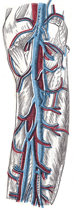
Venae comitantes (paired) of the anterior tibial, posterior tibial and fibular arteries, plus the soleal and gastrocnemius venous sinuses within the calf muscles.
- Course
The tibial and fibular veins join near the popliteus to form the popliteal vein, which runs superficial to the popliteal artery, passes through the adductor hiatus to become the femoral vein (medial to the femoral artery in the femoral triangle), receives the deep femoral vein and the great saphenous vein, and becomes the external iliac vein at the inguinal ligament.
- Tributaries
- Anterior and posterior tibial veins, fibular veins, soleal and gastrocnemius sinuses
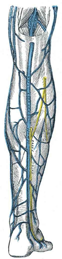
- Small saphenous vein (into the popliteal vein)
- Deep femoral vein and great saphenous vein (into the femoral vein)
- Perforating veins from the superficial system (flow normally superficial → deep)
- Drains into
External iliac vein → common iliac vein → inferior vena cava.
- Relations
The left common iliac vein is crossed by the right common iliac artery (May–Thurner compression → left-sided DVT). Valves are numerous below the knee and absent in the iliac veins.
- Notes
Calf muscle pump: contraction of triceps surae and calcaneal tendon squeezes the deep veins; valves prevent backflow. Immobility abolishes the pump — one limb of Virchow triad (stasis, endothelial injury, hypercoagulability).
Tributaries
Mentioned in
Generated automatically from links in other entries.


Clinical correlations
Deep vein thrombosis PANCE
- Mechanism / lesion
Virchow triad — stasis (immobility, surgery, travel), endothelial injury (trauma, surgery), hypercoagulability (cancer, pregnancy, estrogen, thrombophilia). Usually starts in the calf (soleal sinuses) and propagates proximally.
- Classic presentation
Unilateral calf or leg swelling (calf circumference > 3 cm larger), pain, warmth, pitting edema, dilated superficial veins. Estimate probability with the Wells score: low probability → D-dimer (normal rules out DVT); high probability or positive D-dimer → compression ultrasound. Proximal DVT is treated with anticoagulation (a DOAC for most) for at least 3 months.
Pulmonary embolism PANCE
- Mechanism / lesion
Most PEs arise from proximal (popliteal and above) leg DVTs.
- Classic presentation
Sudden dyspnea, pleuritic chest pain, tachycardia, hypoxia; sinus tachycardia is the most common ECG finding. CT pulmonary angiography for diagnosis.
Phlegmasia cerulea dolens
- Mechanism / lesion
Massive iliofemoral DVT obstructing nearly all venous outflow.
- Classic presentation
A massively swollen, blue, painful leg that can progress to venous gangrene — an emergency (anticoagulation, catheter-directed thrombolysis or thrombectomy).
High-yield PANCE pearls
- Virchow triad: stasis, endothelial injury, hypercoagulability.
- Low pretest probability → D-dimer; high → compression ultrasound.
- Proximal DVT → anticoagulate ≥ 3 months; most PEs come from proximal DVT.
- Left-sided DVT in a young woman → consider May–Thurner (left common iliac vein compression).
- Homans sign is unreliable.
Imaging anatomy
- CT
CT venography for iliac/IVC thrombus; CT pulmonary angiography for PE.
- MRI
MR venography for pelvic veins (pregnancy, May–Thurner).
- Ultrasound
Compression duplex ultrasound — a non-compressible vein with echogenic thrombus is diagnostic; whole-leg or two-point (femoral and popliteal) protocols.
- Key views & landmarks
- Compression ultrasound (femoral and popliteal veins)
Physical exam
- Surface landmarks
The femoral vein lies just medial to the femoral pulse below the inguinal ligament (femoral venous access site); the popliteal vein is superficial to the artery in the popliteal fossa.
- Palpation
Calf tenderness along the deep veins, pitting edema, warmth.
- Strength / function testing
Measure calf circumference 10 cm below the tibial tuberosity on both legs; apply the Wells score.
Self-quiz
A 45-year-old woman returns from a 14-hour flight with a swollen, tender left calf that is 4 cm larger than the right. Her Wells score indicates DVT is likely. What is the best next step?
Correct answer: B
When DVT is likely by Wells score, go straight to compression ultrasound (a normal D-dimer alone is not enough to rule it out in high-probability patients). D-dimer is used first when probability is low.
Show answer
B
When DVT is likely by Wells score, go straight to compression ultrasound (a normal D-dimer alone is not enough to rule it out in high-probability patients). D-dimer is used first when probability is low.
Which is NOT a component of Virchow triad?
Correct answer: D
Virchow triad is venous stasis, endothelial injury and hypercoagulability; systemic arterial hypertension is not part of it.
Show answer
D
Virchow triad is venous stasis, endothelial injury and hypercoagulability; systemic arterial hypertension is not part of it.
A 28-year-old woman has an unprovoked left iliofemoral DVT. Which anatomic relationship may explain its side?
Correct answer: A
In May–Thurner syndrome, the right common iliac artery crosses and compresses the left common iliac vein against the spine, predisposing to left-sided DVT, especially in young women.
Show answer
A
In May–Thurner syndrome, the right common iliac artery crosses and compresses the left common iliac vein against the spine, predisposing to left-sided DVT, especially in young women.
A patient with metastatic cancer develops a massively swollen, painful, bluish left leg with early skin blebs. What is the most likely diagnosis?
Correct answer: C
Phlegmasia cerulea dolens is massive iliofemoral DVT obstructing nearly all venous outflow: a swollen, blue, painful leg that can progress to venous gangrene. It is an emergency treated with anticoagulation and catheter-directed thrombolysis or thrombectomy.
Show answer
C
Phlegmasia cerulea dolens is massive iliofemoral DVT obstructing nearly all venous outflow: a swollen, blue, painful leg that can progress to venous gangrene. It is an emergency treated with anticoagulation and catheter-directed thrombolysis or thrombectomy.
A patient with calf pain has a low pretest probability of DVT by Wells score. What is the most appropriate first test?
Correct answer: B
With low pretest probability, a normal D-dimer rules out DVT. A positive D-dimer, or a high pretest probability, leads to compression ultrasound.
Show answer
B
With low pretest probability, a normal D-dimer rules out DVT. A positive D-dimer, or a high pretest probability, leads to compression ultrasound.