Zygapophysial (facet) joints
Paired synovial joints between the articular processes of adjacent vertebrae. Their orientation determines spinal motion at each level, and facet arthropathy is a common cause of axial back and neck pain.
Core anatomy
- Location
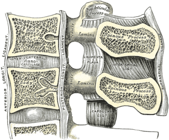
Posterolaterally between the superior articular process of one vertebra and the inferior articular process of the vertebra above, from C2–C3 to L5–S1.
- Classification
Synovial plane joints.
- Articulating surfaces
Articular processes of adjacent vertebrae.
- Capsule
Thin, loose capsule (loosest in the cervical region).
- Ligaments
- Joint capsule; ligamentum flavum anteriorly
- Movements
- Cervical: facets inclined about 45°, allowing flexion–extension, lateral flexion and rotation
- Thoracic: roughly coronal, favoring rotation (limited by ribs)
- Lumbar: roughly sagittal, favoring flexion–extension and limiting rotation
- Stability
Resist shear forces (anterior slippage) and limit rotation, together with the discs and ligaments.
- Innervation
Medial branches of the dorsal rami of the spinal nerves; each joint receives branches from two levels (the level above and at the joint).
- Relations
Form the posterior boundary of the intervertebral foramen; facet hypertrophy narrows the foramen and lateral recess.

Clinical correlations
Facet joint arthropathy PANCE
- Mechanism / lesion
Osteoarthritis of the facets, often with disc degeneration, in older adults.
- Classic presentation
Axial back or neck pain, often worse with extension and rotation and after rest, with poorly localized referral to the buttock or thigh (not below the knee). Diagnosis is confirmed by pain relief after medial branch blocks; radiofrequency ablation of the medial branches is a treatment.
Facet dislocation (cervical)
- Mechanism / lesion
Flexion–distraction injury; unilateral or bilateral locked facets.
- Classic presentation
Neck pain, radiculopathy or cord injury. Bilateral facet dislocation causes marked anterior translation and is highly unstable.
High-yield PANCE pearls
- Facet pain: axial pain worse with extension/rotation; no radiation below the knee.
- Innervated by medial branches of dorsal rami (two levels) → medial branch block/radiofrequency ablation.
- Lumbar facets are sagittal (flexion–extension); thoracic are coronal (rotation).
Imaging anatomy
- X-ray
Facet sclerosis, joint space narrowing and osteophytes on oblique views.
- CT
Best for facet osteoarthritis and fractures or dislocations.
- MRI
Facet effusions, synovial cysts (which can compress roots), and foraminal narrowing.
- Key views & landmarks
- CT or MRI; fluoroscopy-guided diagnostic blocks
Physical exam
- Strength / function testing
Reproduce pain with combined extension and rotation of the spine (facet loading); paraspinal tenderness over the joints. Neurologic exam is typically normal.
Self-quiz
A 66-year-old woman has chronic low back pain without radiation below the knee, worse with extending and twisting the spine. The neurologic exam is normal. What is the most likely source?
Correct answer: B
Axial pain worse with extension and rotation without radicular signs suggests facet arthropathy. Pain relief with medial branch blocks supports the diagnosis.
Show answer
B
Axial pain worse with extension and rotation without radicular signs suggests facet arthropathy. Pain relief with medial branch blocks supports the diagnosis.
Which nerves innervate the zygapophysial joints?
Correct answer: B
Each facet joint is supplied by medial branches of the dorsal rami from two adjacent levels, the target of diagnostic blocks and radiofrequency ablation.
Show answer
B
Each facet joint is supplied by medial branches of the dorsal rami from two adjacent levels, the target of diagnostic blocks and radiofrequency ablation.
The roughly sagittal orientation of lumbar facet joints mainly allows which movement?
Correct answer: B
Lumbar facets lie in a roughly sagittal plane, permitting flexion–extension while limiting rotation.
Show answer
B
Lumbar facets lie in a roughly sagittal plane, permitting flexion–extension while limiting rotation.