Anterior abdominal wall
Layered musculoaponeurotic wall from the costal margin to the pelvis. Its planes and regions are the map for describing abdominal pain, and it is the site of ventral hernias.
Core anatomy
- Location
Between the xiphoid process and costal margins above and the iliac crests, inguinal ligaments and pubic bones below.
- Boundaries
- superior
- Xiphoid process and costal margins (7th–10th costal cartilages)
- inferior
- Iliac crests, inguinal ligaments, pubic crests and pubic symphysis
- lateral
- Mid-axillary lines (continuous with the posterolateral wall)
- Contents
- Layers, superficial to deep: skin; superficial fascia with a fatty layer (Camper) and a membranous layer (Scarpa); external oblique, internal oblique, transversus abdominis; transversalis fascia; extraperitoneal fat; parietal peritoneum
- Rectus abdominis within the rectus sheath on each side of the linea alba
- Thoracoabdominal nerves (T7–T11), subcostal nerve (T12), iliohypogastric and ilioinguinal nerves (L1)
- Superior and inferior epigastric vessels in the rectus sheath; superficial epigastric vessels in the subcutaneous tissue
- Communications
Continuous inferiorly with the inguinal canal, which passes obliquely through its lower part.
- Notes
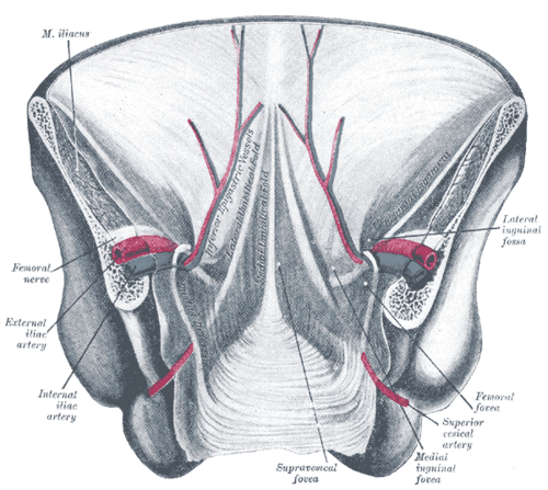
Surface regions. Four quadrants (RUQ, LUQ, RLQ, LLQ) divided by the median plane and the transumbilical plane, or nine regions divided by the midclavicular lines, the subcostal plane and the transtubercular plane (epigastric, umbilical, hypogastric/pubic in the middle; hypochondriac, lumbar/flank, inguinal/iliac on each side). Landmarks: the umbilicus lies at about the L3/L4 disc and the T10 dermatome. The transpyloric plane (L1), halfway between the jugular notch and the pubic symphysis, passes roughly through the pylorus, the fundus of the gallbladder (9th costal cartilage), the neck of the pancreas, the hila of the kidneys and the duodenojejunal flexure. The linea semilunaris marks the lateral edge of the rectus sheath; the arcuate line (roughly one-third of the way from the umbilicus to the pubis) marks where the posterior rectus sheath ends. verify

Clinical correlations
Ventral hernias (umbilical, epigastric, incisional, Spigelian) PANCE
- Mechanism / lesion
Protrusion through a weakness in the wall. Umbilical: common in infants (most close spontaneously in early childhood) and in adults with raised intra-abdominal pressure (obesity, pregnancy, ascites). Epigastric: through the linea alba above the umbilicus. Incisional: through a surgical scar. Spigelian: through the semilunar line, usually near the arcuate line.
- Classic presentation
A bulge that enlarges with coughing or straining. Pain, tenderness, irreducibility or signs of obstruction suggest incarceration or strangulation, a surgical emergency.
Referred and localized abdominal pain PANCE
- Mechanism / lesion
Visceral pain (autonomic afferents) is poorly localized to the midline by embryologic origin: foregut → epigastric, midgut → periumbilical, hindgut → suprapubic. Parietal peritoneal pain (somatic nerves of the wall) is sharp and well localized.
- Classic presentation
For example, appendicitis begins as periumbilical pain (visceral), then localizes to the RLQ once the parietal peritoneum is inflamed.
Abdominal wall signs of intra-abdominal bleeding PANCE
- Mechanism / lesion
Blood tracking from the retroperitoneum or peritoneal cavity into the subcutaneous tissue.
- Classic presentation
Cullen sign (periumbilical ecchymosis) and Grey Turner sign (flank ecchymosis), e.g. in severe hemorrhagic pancreatitis or ruptured ectopic pregnancy. Caput medusae (dilated periumbilical veins) indicates portal hypertension.
High-yield PANCE pearls
- Visceral pain: foregut → epigastrium, midgut → periumbilical, hindgut → suprapubic.
- Umbilicus = T10 dermatome, about L3/L4.
- Cullen (periumbilical) and Grey Turner (flank) ecchymosis → hemorrhagic pancreatitis, ruptured ectopic.
- Carnett sign separates abdominal wall pain from visceral pain.
- Incarcerated hernia with tenderness, erythema or obstructive symptoms → urgent surgical evaluation.
Imaging anatomy
- X-ray
Limited. Free intraperitoneal air under the diaphragm on an upright CXR; bowel gas within a hernia sac.
- CT
CT defines hernia contents, the neck of the defect, incarceration/strangulation, abdominal wall masses, hematomas and abscesses.
- Ultrasound
Dynamic ultrasound with Valsalva identifies ventral hernias and their contents at the bedside, distinguishes hernias from lipomas or hematomas, and guides abdominal wall blocks (e.g. the TAP block in the plane between internal oblique and transversus abdominis).
- Key views & landmarks
- CT abdomen/pelvis with contrast
- Ultrasound over the bulge at rest and with Valsalva
Physical exam
- Surface landmarks
Xiphoid, costal margins, umbilicus, anterior superior iliac spines, pubic tubercles, linea alba, linea semilunaris.
- Palpation
Order of the abdominal exam: inspection → auscultation → percussion → palpation (auscultate before palpation so bowel sounds aren't altered). Palpate lightly then deeply, starting away from the painful area. Check for hernias with the patient coughing or lifting the head.
- Strength / function testing
Look for guarding, rigidity and rebound tenderness (peritonitis). Tensing the wall (head or legs raised) makes diastasis recti and ventral hernias more obvious.
Carnett sign
- Technique
Locate the point of maximal tenderness, then have the patient tense the abdominal muscles (lift the head and shoulders or both legs) while you press again.
- Positive test
Tenderness stays the same or increases with the muscles tensed.
- Significance
Suggests an abdominal wall source (e.g. nerve entrapment, hernia, hematoma). Visceral tenderness usually decreases because the tense muscles shield the viscera.
Self-quiz
A 19-year-old man has vague periumbilical pain that, over 12 hours, becomes sharp and localized to the right lower quadrant. What explains the shift?
Correct answer: B
Early appendiceal distension stimulates visceral afferents entering at about T10, giving poorly localized periumbilical pain (midgut). When inflammation reaches the parietal peritoneum (somatic innervation), the pain becomes sharp and localized to the RLQ.
Show answer
B
Early appendiceal distension stimulates visceral afferents entering at about T10, giving poorly localized periumbilical pain (midgut). When inflammation reaches the parietal peritoneum (somatic innervation), the pain becomes sharp and localized to the RLQ.
Which dermatome corresponds to the umbilicus?
Correct answer: C
The umbilicus lies in the T10 dermatome. T4 is the nipple; L1 is the inguinal region.
Show answer
C
The umbilicus lies in the T10 dermatome. T4 is the nipple; L1 is the inguinal region.
A patient has point tenderness in the right lower abdominal wall. When she lifts her head and shoulders off the table, the tenderness increases. What does this suggest?
Correct answer: B
A positive Carnett sign (tenderness unchanged or worse with the wall tensed) points to an abdominal wall source such as anterior cutaneous nerve entrapment or a hernia. Visceral pain typically lessens.
Show answer
B
A positive Carnett sign (tenderness unchanged or worse with the wall tensed) points to an abdominal wall source such as anterior cutaneous nerve entrapment or a hernia. Visceral pain typically lessens.
A patient with severe hemorrhagic pancreatitis develops bruising around the umbilicus. What is this sign called?
Correct answer: B
Cullen sign is periumbilical ecchymosis; Grey Turner sign is flank ecchymosis. Both reflect blood tracking from the retroperitoneum or peritoneal cavity, e.g. in hemorrhagic pancreatitis or ruptured ectopic pregnancy. Caput medusae is dilated periumbilical veins in portal hypertension.
Show answer
B
Cullen sign is periumbilical ecchymosis; Grey Turner sign is flank ecchymosis. Both reflect blood tracking from the retroperitoneum or peritoneal cavity, e.g. in hemorrhagic pancreatitis or ruptured ectopic pregnancy. Caput medusae is dilated periumbilical veins in portal hypertension.
A hernia protrudes through the lateral edge of the rectus sheath in the lower abdomen, near the arcuate line. What type of hernia is it?
Correct answer: E
A Spigelian hernia passes through the linea semilunaris (the lateral edge of the rectus sheath), usually near the arcuate line. Epigastric hernias pass through the linea alba above the umbilicus.
Show answer
E
A Spigelian hernia passes through the linea semilunaris (the lateral edge of the rectus sheath), usually near the arcuate line. Epigastric hernias pass through the linea alba above the umbilicus.