External and middle ear
The ear canal, eardrum and air-filled tympanic cavity with its three ossicles, connected to the nasopharynx by the pharyngotympanic tube. Otitis externa, otitis media and conductive hearing loss are the key topics.
Core anatomy
- Location
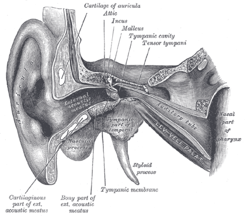
External ear: auricle and external acoustic meatus (about 2.5 cm, S-shaped). Middle ear: tympanic cavity within the petrous temporal bone, between the tympanic membrane and the inner ear.
- Parts
- Tympanic membrane: pearly gray, cone-shaped; the handle of the malleus is attached (umbo at its tip); the cone of light points anteroinferiorly; pars flaccida (above) and pars tensa
- Ossicles: malleus → incus → stapes (footplate in the oval window); they amplify sound
- Muscles: tensor tympani (V3) and stapedius (CN VII) — dampen loud sounds (stapedius paralysis → hyperacusis)
- Pharyngotympanic (Eustachian) tube: equalizes pressure with the nasopharynx; shorter, wider and more horizontal in children
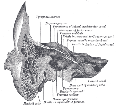
- Mastoid antrum and air cells: posterior communication — route for spread of infection
- Walls: roof (tegmen tympani; middle cranial fossa above); floor (internal jugular vein); medial wall (promontory, oval and round windows, facial canal); posterior (mastoid)
- Relations
The facial nerve runs in its bony canal in the medial and posterior walls — at risk with middle ear disease and surgery.
- Arterial supply
External canal — posterior auricular and superficial temporal arteries; middle ear — tympanic branches of the maxillary and other external carotid artery branches.
- Venous drainage
Pterygoid plexus and superior petrosal sinus.
- Lymphatic drainage
Preauricular, mastoid and deep cervical nodes.
- Innervation
External canal and outer drum — auriculotemporal (V3), vagus (Arnold nerve — cough reflex when the canal is touched), facial and great auricular nerves. Middle ear mucosa — tympanic branch of CN IX. The chorda tympani (CN VII, taste) crosses the medial surface of the drum.
Mentioned in
Generated automatically from links in other entries.


Clinical correlations
Acute otitis media PANCE
- Mechanism / lesion
Eustachian tube dysfunction after a viral URI; S. pneumoniae, nontypeable H. influenzae, M. catarrhalis. Most common in children (short, horizontal tube).
- Classic presentation
Ear pain, fever, irritability; bulging, erythematous, opaque tympanic membrane with reduced mobility on pneumatic otoscopy. First-line antibiotic is high-dose amoxicillin (amoxicillin-clavulanate if recent amoxicillin or conjunctivitis); watchful waiting is an option for some older children with mild disease. Complications: perforation, mastoiditis, facial palsy, meningitis.
Otitis externa ("swimmer's ear") PANCE
- Mechanism / lesion
Infection of the canal skin, usually Pseudomonas or S. aureus, after moisture or trauma. Malignant (necrotizing) otitis externa — Pseudomonas osteomyelitis of the skull base in diabetic or immunocompromised older adults.
- Classic presentation
Ear pain with pain on pulling the auricle or pressing the tragus, canal edema and discharge; the drum is normal if seen. Treat with topical antibiotic (fluoroquinolone) drops ± steroid; malignant OE needs IV antipseudomonal antibiotics and imaging.
Otitis media with effusion and cholesteatoma
- Mechanism / lesion
Effusion: fluid behind an intact drum after AOM or with Eustachian tube dysfunction. Cholesteatoma: keratinizing squamous epithelium in the middle ear eroding bone.
- Classic presentation
Effusion — conductive hearing loss, dull retracted drum with air-fluid levels; tympanostomy tubes if persistent with hearing loss. Cholesteatoma — painless foul discharge, white mass (often pars flaccida), hearing loss; surgical removal.
High-yield PANCE pearls
- AOM: bulging drum, reduced mobility → high-dose amoxicillin.
- Pain with tragal pressure or auricle traction → otitis externa (Pseudomonas) → topical drops.
- Elderly diabetic with severe ear pain, granulation tissue in the canal → malignant OE.
- Middle ear disease → conductive loss: Weber lateralizes to the affected ear; Rinne BC > AC.
- Post-auricular swelling, protruding ear after AOM → mastoiditis.
Imaging anatomy
- CT
Temporal bone CT for mastoiditis, cholesteatoma, trauma and malignant otitis externa (bone erosion).
- MRI
Intracranial complications (abscess, venous sinus thrombosis); MRI diffusion-weighted imaging for recurrent cholesteatoma.
- Key views & landmarks
- CT temporal bones (thin section)
Physical exam
- Surface landmarks
Tragus, mastoid process, external acoustic meatus.
- Palpation
Pull the auricle and press the tragus (pain = otitis externa); palpate the mastoid for tenderness and swelling.
- Strength / function testing
Otoscopy: pull the auricle up and back in adults (down and back in young children). Look for the cone of light, handle of malleus, color, position (bulging/retracted), perforation, and use pneumatic otoscopy for mobility. Hearing: whispered voice, Weber and Rinne tuning-fork tests (512 Hz).
Rinne test
- Technique
Place a vibrating 512-Hz tuning fork on the mastoid (bone conduction), then beside the ear canal (air conduction); ask which is louder or longer.
- Positive test
"Negative" Rinne — bone conduction louder than air conduction.
- Significance
Conductive hearing loss in that ear (normal and sensorineural loss show AC > BC).
Weber test
- Technique
Place the vibrating fork on the midline of the forehead or vertex.
- Positive test
Sound lateralizes to one ear.
- Significance
Lateralizes toward the ear with conductive loss, or away from the ear with sensorineural loss.
Self-quiz
A 2-year-old has fever and pulls at his right ear after a cold. Otoscopy shows a bulging, red, opaque tympanic membrane that does not move with insufflation. He has no penicillin allergy and no recent antibiotics. What is the best treatment?
Correct answer: A
A bulging, immobile drum confirms acute otitis media. First-line therapy is high-dose amoxicillin. Topical fluoroquinolones treat otitis externa; tubes are for recurrent AOM or persistent effusion.
Show answer
A
A bulging, immobile drum confirms acute otitis media. First-line therapy is high-dose amoxicillin. Topical fluoroquinolones treat otitis externa; tubes are for recurrent AOM or persistent effusion.
A patient has left-sided hearing loss. Weber lateralizes to the left, and Rinne on the left shows bone conduction louder than air conduction. What type of hearing loss is present?
Correct answer: B
In conductive loss, Weber lateralizes to the affected ear and Rinne shows BC > AC on that side (e.g. cerumen, effusion, otosclerosis). In sensorineural loss, Weber lateralizes to the good ear and AC > BC.
Show answer
B
In conductive loss, Weber lateralizes to the affected ear and Rinne shows BC > AC on that side (e.g. cerumen, effusion, otosclerosis). In sensorineural loss, Weber lateralizes to the good ear and AC > BC.
A swimmer has ear pain that worsens when the examiner pulls on the auricle or presses the tragus. What is the most likely diagnosis?
Correct answer: C
Otitis externa (usually Pseudomonas or S. aureus) causes pain on auricle traction or tragal pressure with canal edema and discharge. Treat with topical antibiotic drops.
Show answer
C
Otitis externa (usually Pseudomonas or S. aureus) causes pain on auricle traction or tragal pressure with canal edema and discharge. Treat with topical antibiotic drops.
A 74-year-old man with poorly controlled diabetes has severe ear pain and granulation tissue in the ear canal. Which organism and condition are most likely?
Correct answer: E
Malignant otitis externa is Pseudomonas osteomyelitis of the skull base in diabetic or immunocompromised older adults. It needs IV antipseudomonal antibiotics and imaging.
Show answer
E
Malignant otitis externa is Pseudomonas osteomyelitis of the skull base in diabetic or immunocompromised older adults. It needs IV antipseudomonal antibiotics and imaging.
Paralysis of which middle ear muscle causes hyperacusis in a facial nerve palsy?
Correct answer: A
Stapedius (CN VII) dampens loud sounds; its paralysis causes hyperacusis. Tensor tympani is supplied by V3.
Show answer
A
Stapedius (CN VII) dampens loud sounds; its paralysis causes hyperacusis. Tensor tympani is supplied by V3.