Suprarenal gland
Paired retroperitoneal endocrine glands on the superomedial poles of the kidneys. The cortex makes steroids and the medulla makes catecholamines; pheochromocytoma, Cushing, Conn and Addison are the classic disorders.
Core anatomy
- Location
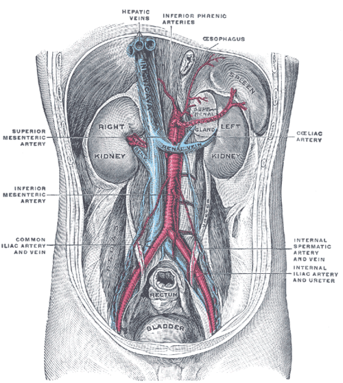
Retroperitoneal, superomedial to each kidney, within the renal (Gerota) fascia but in a separate compartment. Right: pyramidal, behind the IVC. Left: crescentic (semilunar), near the aorta, behind the stomach and pancreas.
- Parts
- Cortex (mesoderm), outer to inner: zona glomerulosa (aldosterone), zona fasciculata (cortisol), zona reticularis (androgens). Mnemonic: "salt, sugar, sex" (GFR)
- Medulla (neural crest): chromaffin cells secreting epinephrine and norepinephrine. It is innervated directly by preganglionic sympathetic fibers (it functions as a modified sympathetic ganglion)
- Relations
Right gland lies behind the IVC and near the bare area of the liver; left gland near the splenic vessels, pancreas and stomach. Both lie on the crura of the diaphragm.
- Arterial supply
Superior suprarenal (from the inferior phrenic), middle suprarenal (from the aorta), inferior suprarenal (from the renal) arteries.
- Venous drainage
Single suprarenal vein on each side. The right drains directly into the IVC (very short, a hazard in adrenalectomy); the left drains into the left renal vein.
- Innervation
Celiac plexus and thoracic splanchnic nerves (preganglionic sympathetic fibers to the medulla).

Clinical correlations
Pheochromocytoma PANCE
- Mechanism / lesion
Catecholamine-secreting tumor of the medulla (chromaffin cells). Associated with MEN 2, von Hippel–Lindau and NF1.
- Classic presentation
Episodic headache, sweating and palpitations (tachycardia) with hypertension (paroxysmal or sustained). Screen with plasma free or 24-hour urine fractionated metanephrines, then image. Before surgery, give alpha blockade first (phenoxybenzamine), then beta blockade, to avoid unopposed alpha stimulation.
Primary hyperaldosteronism (Conn syndrome) PANCE
- Mechanism / lesion
Aldosterone-producing adenoma or bilateral adrenal hyperplasia.
- Classic presentation
Hypertension with hypokalemia (often), metabolic alkalosis, low renin. Screen with the aldosterone-to-renin ratio.
Adrenal insufficiency (Addison disease) and adrenal crisis PANCE
- Mechanism / lesion
Primary adrenal failure, most often autoimmune in developed countries (also TB, hemorrhage, e.g. Waterhouse–Friderichsen in meningococcemia, metastases).
- Classic presentation
Fatigue, weight loss, hyperpigmentation (raised ACTH), hypotension, hyponatremia, hyperkalemia, hypoglycemia. Adrenal crisis: shock; treat immediately with IV hydrocortisone and fluids.
Adrenal incidentaloma and Cushing syndrome
- Mechanism / lesion
Adrenal masses are often found on imaging done for other reasons; some secrete cortisol (Cushing), aldosterone or catecholamines, and a few are malignant.
- Classic presentation
Every incidentaloma is evaluated for hormone excess (cortisol, catecholamines, aldosterone if hypertensive) and malignancy (size, imaging features). Larger masses (commonly more than 4 cm) raise concern.
High-yield PANCE pearls
- Cortex GFR: glomerulosa (aldosterone), fasciculata (cortisol), reticularis (androgens).
- Pheochromocytoma: headache + sweating + tachycardia + HTN; metanephrines; alpha before beta blockade.
- Conn: HTN + hypokalemia; aldosterone:renin ratio.
- Addison: hyperpigmentation, hyponatremia, hyperkalemia; crisis → IV hydrocortisone.
- Right suprarenal vein → IVC; left → left renal vein.
Imaging anatomy
- CT
Adrenal-protocol CT: an unenhanced attenuation of 10 HU or less indicates a lipid-rich (benign) adenoma; contrast washout characteristics help with others. Pheochromocytomas are usually enhancing and higher in attenuation.
- MRI
Chemical-shift MRI shows signal loss in lipid-rich adenomas; pheochromocytomas are classically bright on T2.
- Ultrasound
Limited for the adrenals (large masses may be seen); not a primary tool.
- Key views & landmarks
- Adrenal-protocol CT (unenhanced + contrast washout)
- Functional imaging (e.g. MIBG or PET) for pheochromocytoma localization
Physical exam
- Strength / function testing
Blood pressure (including paroxysms), orthostatic changes (insufficiency), hyperpigmentation (palmar creases, buccal mucosa), Cushingoid features (central obesity, purple striae, proximal weakness).
Self-quiz
A 38-year-old woman has episodes of pounding headache, sweating and palpitations, with BP 210/120 during an episode. What is the best initial test?
Correct answer: B
The triad of headache, sweating and tachycardia with hypertension suggests pheochromocytoma. Screen biochemically with plasma free (or urine fractionated) metanephrines before imaging.
Show answer
B
The triad of headache, sweating and tachycardia with hypertension suggests pheochromocytoma. Screen biochemically with plasma free (or urine fractionated) metanephrines before imaging.
Before surgical removal of a pheochromocytoma, which medication should be started first?
Correct answer: B
Alpha blockade (phenoxybenzamine) comes first. Starting a beta blocker first leaves alpha-mediated vasoconstriction unopposed and can precipitate a hypertensive crisis.
Show answer
B
Alpha blockade (phenoxybenzamine) comes first. Starting a beta blocker first leaves alpha-mediated vasoconstriction unopposed and can precipitate a hypertensive crisis.
The right suprarenal vein drains into which vessel?
Correct answer: B
The right suprarenal vein is very short and drains directly into the IVC; the left drains into the left renal vein.
Show answer
B
The right suprarenal vein is very short and drains directly into the IVC; the left drains into the left renal vein.
A patient has fatigue, hyperpigmented palmar creases, BP 88/56, Na 128 and K 5.9. What is the most likely diagnosis?
Correct answer: B
Hyperpigmentation (high ACTH) with hyponatremia and hyperkalemia (loss of aldosterone) indicates primary adrenal insufficiency. Secondary insufficiency lacks hyperpigmentation and hyperkalemia.
Show answer
B
Hyperpigmentation (high ACTH) with hyponatremia and hyperkalemia (loss of aldosterone) indicates primary adrenal insufficiency. Secondary insufficiency lacks hyperpigmentation and hyperkalemia.
A 45-year-old with resistant hypertension has a serum potassium of 3.0 mEq/L and metabolic alkalosis. What is the best initial screening test for the most likely diagnosis?
Correct answer: A
Primary hyperaldosteronism (Conn syndrome) causes hypertension with hypokalemia, metabolic alkalosis and low renin. Screen with the aldosterone-to-renin ratio. Metanephrines screen for pheochromocytoma.
Show answer
A
Primary hyperaldosteronism (Conn syndrome) causes hypertension with hypokalemia, metabolic alkalosis and low renin. Screen with the aldosterone-to-renin ratio. Metanephrines screen for pheochromocytoma.