Pericardium
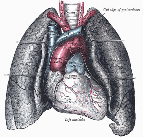
Double-walled fibroserous sac enclosing the heart. Pericarditis, effusion and tamponade are its key clinical problems.
Core anatomy
- Location
Middle mediastinum, enclosing the heart and the roots of the great vessels. Fused to the central tendon of the diaphragm below.
- Parts
- Fibrous pericardium: tough outer layer, continuous with the adventitia of the great vessels, attached to the diaphragm and sternum; limits acute dilation
- Serous pericardium, parietal layer: lines the fibrous pericardium
- Serous pericardium, visceral layer (epicardium): covers the heart surface
- Pericardial cavity: potential space between the parietal and visceral serous layers, normally containing a small amount of lubricating fluid
- Transverse pericardial sinus: passage posterior to the ascending aorta and pulmonary trunk, anterior to the SVC. Surgeons pass a finger or ligature through it to clamp the great arteries during cardiac surgery
- Oblique pericardial sinus: blind recess behind the left atrium, bounded by the pulmonary veins and IVC
- Relations
The phrenic nerve and pericardiacophrenic vessels descend on each side between the fibrous pericardium and the mediastinal pleura.
- Arterial supply
Pericardiacophrenic artery (from the internal thoracic artery) mainly, plus musculophrenic, bronchial, esophageal and superior phrenic branches.
- Innervation
Phrenic nerves (fibrous and parietal layers): sensory, so pain from pericarditis can be referred to the shoulder or trapezius ridge (C3–C5). The visceral layer (epicardium) is insensitive to pain. Also vagus and sympathetic fibers.
Swipe sideways to see the whole diagram →

Clinical correlations
Acute pericarditis PANCE
- Mechanism / lesion
Inflammation of the pericardium, most often idiopathic or viral. Other causes include uremia, post-MI (including Dressler syndrome), autoimmune disease, malignancy and TB.
- Classic presentation
Sharp, pleuritic chest pain that is better sitting up and leaning forward, worse lying flat, and may radiate to the trapezius ridge. Pericardial friction rub. ECG: diffuse concave ST elevation with PR depression.
Cardiac tamponade PANCE
- Mechanism / lesion
Fluid (blood, effusion) accumulating in the pericardial cavity faster than the fibrous pericardium can stretch, compressing the heart and impairing filling.
- Classic presentation
Beck's triad: hypotension, distended neck veins (JVD), muffled heart sounds. Also tachycardia, pulsus paradoxus (a drop in systolic BP of more than 10 mmHg on inspiration), and electrical alternans on ECG. Treat with pericardiocentesis (or surgical drainage).
Constrictive pericarditis
- Mechanism / lesion
A thickened, fibrotic (sometimes calcified) pericardium restricts diastolic filling. Causes include prior cardiac surgery, radiation, TB, and previous pericarditis.
- Classic presentation
Right heart failure signs (JVD, edema, ascites), Kussmaul sign (JVP rises on inspiration), and a pericardial knock in early diastole.
High-yield PANCE pearls
- Pericarditis: pain relieved leaning forward, friction rub, diffuse ST elevation + PR depression.
- Tamponade: Beck's triad (hypotension, JVD, muffled heart sounds) + pulsus paradoxus + electrical alternans.
- Constrictive pericarditis: Kussmaul sign, pericardial knock, calcified pericardium.
- Pericardial pain can refer to the trapezius ridge via the phrenic nerve.
Imaging anatomy
- X-ray
A large effusion gives a globular "water bottle" heart with clear lungs. Pericardial calcification suggests constrictive pericarditis. A normal CXR doesn't exclude tamponade (it depends on the rate of accumulation).
- CT
Pericardial thickening (more than about 4 mm suggests constriction), calcification, and effusion characteristics. verify
- MRI
Pericardial thickening, inflammation (late enhancement), and constrictive physiology (septal bounce on real-time cine).
- Ultrasound
Echocardiography is the key test. Effusion appears as an anechoic space around the heart. Tamponade signs: right atrial systolic collapse and right ventricular diastolic collapse, a plethoric IVC without respiratory variation, and exaggerated respiratory variation in mitral and tricuspid inflow. The subcostal view in the FAST exam screens for pericardial fluid in trauma. Echo guides pericardiocentesis.
- Key views & landmarks
- Subcostal (FAST) and parasternal long axis views
- ECG: PR depression, diffuse ST elevation (pericarditis); low voltage and electrical alternans (effusion/tamponade)
Physical exam
- Surface landmarks
Subxiphoid pericardiocentesis (the classic approach): the needle enters between the xiphoid process and the left costal margin and is directed toward the left shoulder at about 30–45° to the skin, ideally under echo guidance. verify
- Palpation
The apical impulse may be impalpable with a large effusion.
- Strength / function testing
Auscultate for a friction rub (scratchy, with up to three components; best heard at the left lower sternal border with the patient leaning forward and holding the breath). Measure pulsus paradoxus with a manual cuff. Assess the JVP and Kussmaul sign.
Self-quiz
A 30-year-old man with a recent viral illness has sharp chest pain that improves when he sits up and leans forward. A scratchy sound is heard at the left sternal border. ECG shows diffuse ST elevation with PR depression. What is the diagnosis?
Correct answer: B
Positional pleuritic pain, a friction rub, and diffuse ST elevation with PR depression are classic for acute pericarditis. An MI would show territorial ST elevation with reciprocal changes.
Show answer
B
Positional pleuritic pain, a friction rub, and diffuse ST elevation with PR depression are classic for acute pericarditis. An MI would show territorial ST elevation with reciprocal changes.
After a stab wound to the chest, a patient has BP 78/50, distended neck veins and distant heart sounds. Breath sounds are equal bilaterally. What is the most appropriate immediate intervention?
Correct answer: B
Beck's triad with equal breath sounds indicates cardiac tamponade. Relieve it with pericardiocentesis as a temporizing measure, or surgical drainage (definitive in trauma). Tension pneumothorax would have absent breath sounds on one side.
Show answer
B
Beck's triad with equal breath sounds indicates cardiac tamponade. Relieve it with pericardiocentesis as a temporizing measure, or surgical drainage (definitive in trauma). Tension pneumothorax would have absent breath sounds on one side.
During cardiac surgery, the surgeon passes a finger posterior to the ascending aorta and pulmonary trunk to place a clamp. Which space is used?
Correct answer: B
The transverse pericardial sinus lies posterior to the ascending aorta and pulmonary trunk and anterior to the SVC, allowing the great arteries to be isolated.
Show answer
B
The transverse pericardial sinus lies posterior to the ascending aorta and pulmonary trunk and anterior to the SVC, allowing the great arteries to be isolated.
Years after mediastinal radiation, a patient has edema, ascites and distended neck veins. The JVP rises on inspiration, and there is an early diastolic knock. What is the most likely diagnosis?
Correct answer: C
Constrictive pericarditis (after radiation, cardiac surgery, TB or prior pericarditis) restricts diastolic filling, causing right heart failure signs, a Kussmaul sign (JVP rising on inspiration) and a pericardial knock.
Show answer
C
Constrictive pericarditis (after radiation, cardiac surgery, TB or prior pericarditis) restricts diastolic filling, causing right heart failure signs, a Kussmaul sign (JVP rising on inspiration) and a pericardial knock.
In cardiac tamponade, what does pulsus paradoxus mean?
Correct answer: B
Pulsus paradoxus is an exaggerated fall in systolic BP (more than 10 mmHg) during inspiration. With Beck's triad (hypotension, JVD, muffled heart sounds) and electrical alternans, it supports tamponade.
Show answer
B
Pulsus paradoxus is an exaggerated fall in systolic BP (more than 10 mmHg) during inspiration. With Beck's triad (hypotension, JVD, muffled heart sounds) and electrical alternans, it supports tamponade.