Axillary artery
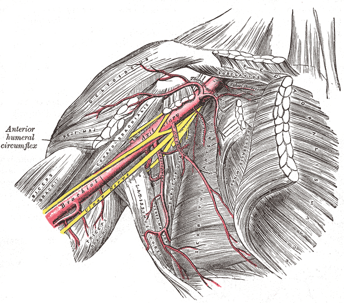
Continuation of the subclavian artery through the axilla, divided into three parts by pectoralis minor, with six named branches.
Core anatomy
- Location
Axilla, from the lateral border of the first rib to the inferior border of teres major.
- Origin
Continuation of the subclavian artery at the lateral border of the first rib.
- Course
Descends through the axilla within the axillary sheath with the cords of the brachial plexus. Divided by pectoralis minor into three parts:
- First part: medial to pectoralis minor. One branch. - Second part: posterior to pectoralis minor. Two branches. The cords are named by their relation to this part. - Third part: lateral to pectoralis minor. Three branches.
- Branches
- 1st part: superior thoracic artery
- 2nd part: thoracoacromial artery (clavicular, acromial, deltoid, pectoral branches); lateral thoracic artery
- 3rd part: subscapular artery (→ circumflex scapular and thoracodorsal arteries); anterior circumflex humeral artery; posterior circumflex humeral artery (with the axillary nerve through the quadrangular space)
- Mnemonic (in order): Screw The Lawyer Save A Patient
- Supplies
Axillary walls, pectoral region, lateral thoracic wall (including the lateral breast), scapular muscles, and the shoulder joint and proximal humerus.
- Anastomoses
Scapular anastomosis around the scapula: branches of the subclavian artery (suprascapular artery; dorsal scapular artery or deep branch of the transverse cervical artery) connect with branches of the axillary artery (circumflex scapular, thoracodorsal). If the axillary artery is occluded between the thyrocervical trunk and the subscapular artery, flow can reverse through this network to reach the third part. Around the surgical neck, the anterior and posterior circumflex humeral arteries anastomose.
- Termination
Becomes the brachial artery at the inferior border of teres major.
- Relations
The axillary vein lies anteromedial to the artery. The cords of the brachial plexus surround the second part (lateral, medial and posterior to it), and their branches surround the third part. The median nerve forms anterior to the third part.
Branches
Structures supplied
Found in
Mentioned in
Generated automatically from links in other entries.

Clinical correlations
Axillary artery injury
- Mechanism / lesion
Penetrating trauma, or blunt injury with anterior shoulder dislocation or proximal humerus fracture (older patients with atherosclerotic, less compliant vessels are at higher risk).
- Classic presentation
Expanding axillary hematoma, diminished or absent distal pulses, and signs of limb ischemia. Associated brachial plexus injury is common because of their close relationship.
Collateral circulation after proximal occlusion PANCE
- Mechanism / lesion
Slow occlusion or ligation of the axillary artery between the thyrocervical trunk and subscapular artery.
- Classic presentation
The limb may remain viable through the scapular anastomosis. Occlusion distal to the subscapular artery has much poorer collateral flow.
Posterior circumflex humeral artery injury PANCE
- Mechanism / lesion
Surgical neck fracture of the humerus (it runs with the axillary nerve around the surgical neck).
- Classic presentation
Bleeding or hematoma with a proximal humerus fracture; accompanies axillary nerve injury.
High-yield PANCE pearls
- Pectoralis minor divides the axillary artery into 3 parts with 1, 2 and 3 branches.
- Axillary artery becomes brachial at the inferior border of teres major.
- Scapular anastomosis protects the limb if the artery is ligated proximal to the subscapular artery.
- Posterior circumflex humeral artery + axillary nerve → quadrangular space and surgical neck.
Imaging anatomy
- X-ray
Not visible. Radiographs show dislocations or fractures that put it at risk.
- CT
CT angiography is the main modality in trauma or suspected occlusion. It shows extravasation, pseudoaneurysm, dissection or occlusion and the collateral network.
- MRI
MR angiography is an alternative for non-emergent vascular evaluation.
- Ultrasound
Duplex ultrasound in the axilla shows flow, stenosis, thrombosis and pseudoaneurysm. The artery is the landmark for ultrasound-guided axillary brachial plexus block, with the terminal nerves arranged around it.
- Key views & landmarks
- CT angiography of the upper extremity
- Duplex ultrasound, axillary approach
Physical exam
- Surface landmarks
Pulse in the axilla against the humerus along the lateral wall of the axilla, with the arm abducted.
- Palpation
Palpate high in the axilla against the humerus, lateral to the axillary hair-bearing area, with the arm abducted about 90°.
- Strength / function testing
In shoulder trauma, compare radial and brachial pulses and capillary refill with the other side. Check the brachial plexus as well.
Self-quiz
Which branch of the axillary artery arises from its second part?
Correct answer: C
The second part (posterior to pectoralis minor) gives the thoracoacromial and lateral thoracic arteries. The superior thoracic artery comes from the first part; the subscapular and both circumflex humeral arteries from the third.
Show answer
C
The second part (posterior to pectoralis minor) gives the thoracoacromial and lateral thoracic arteries. The superior thoracic artery comes from the first part; the subscapular and both circumflex humeral arteries from the third.
A surgeon must ligate the axillary artery. Ligation at which site is most likely to preserve distal perfusion through collateral flow?
Correct answer: B
Between the thyrocervical trunk (subclavian) and the subscapular artery (axillary), the scapular anastomosis can reverse flow around the scapula into the subscapular artery and on to the third part.
Show answer
B
Between the thyrocervical trunk (subclavian) and the subscapular artery (axillary), the scapular anastomosis can reverse flow around the scapula into the subscapular artery and on to the third part.
The axillary artery becomes the brachial artery at which landmark?
Correct answer: C
The subclavian becomes axillary at the lateral border of the first rib, and the axillary becomes brachial at the inferior border of teres major.
Show answer
C
The subclavian becomes axillary at the lateral border of the first rib, and the axillary becomes brachial at the inferior border of teres major.
Which branch arises from the third part of the axillary artery (lateral to pectoralis minor)?
Correct answer: D
The third part has three branches: the subscapular, anterior circumflex humeral and posterior circumflex humeral arteries. The superior thoracic comes from the first part; the thoracoacromial and lateral thoracic from the second. The profunda brachii is a branch of the brachial artery.
Show answer
D
The third part has three branches: the subscapular, anterior circumflex humeral and posterior circumflex humeral arteries. The superior thoracic comes from the first part; the thoracoacromial and lateral thoracic from the second. The profunda brachii is a branch of the brachial artery.
Pectoralis minor crosses which part of the axillary artery, and how many branches does that part give off?
Correct answer: A
Pectoralis minor lies anterior to the second part, which gives two branches (thoracoacromial and lateral thoracic). The first part (medial to the muscle) has one branch and the third part (lateral to it) has three: the "1, 2, 3" rule. The cords of the brachial plexus are named by their relation to the second part.
Show answer
A
Pectoralis minor lies anterior to the second part, which gives two branches (thoracoacromial and lateral thoracic). The first part (medial to the muscle) has one branch and the third part (lateral to it) has three: the "1, 2, 3" rule. The cords of the brachial plexus are named by their relation to the second part.