Radius
Lateral bone of the forearm. It rotates around the ulna in pronation and supination, and its distal end is one of the most commonly fractured sites in the body.
Core anatomy
- Location
Lateral (thumb side) of the forearm; narrow proximally, broad distally.
- Classification
Long bone.
- Key features & landmarks
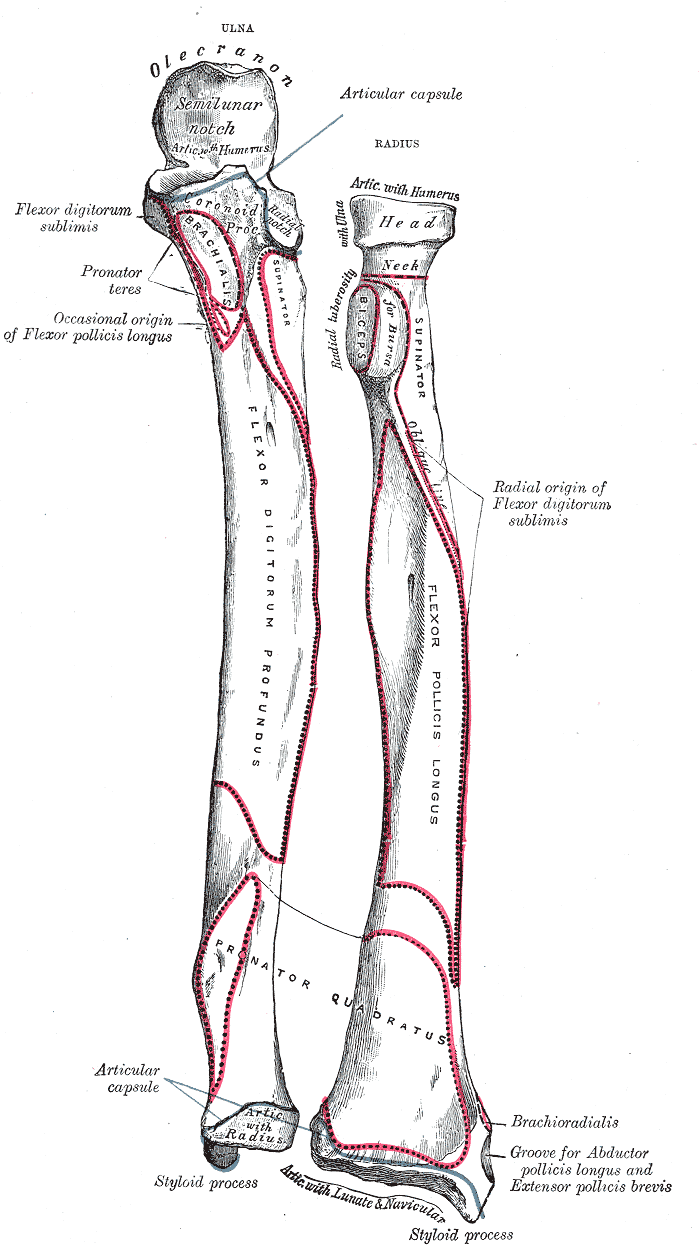
- Head: disc-shaped; articulates with the capitulum of the humerus and the radial notch of the ulna; encircled by the anular ligament
- Neck and radial tuberosity (insertion of biceps brachii)
- Pronator tuberosity: roughened area mid-lateral shaft (insertion of pronator teres)
- Interosseous border: attachment of the interosseous membrane
- Ulnar notch (distal radioulnar joint) and carpal articular surface (scaphoid and lunate)
- Radial styloid process: extends about 1 cm further distally than the ulnar styloid
- Dorsal (Lister) tubercle: the extensor pollicis longus tendon turns around it
- Relations
Proximal and distal radioulnar joints allow the radius to rotate around the ulna. The radial artery is palpable anterior to its distal end, lateral to the FCR tendon.
Muscle attachments
- Biceps brachii — insertion
- Pronator teres — insertion
Joints
Mentioned in
Generated automatically from links in other entries.

Clinical correlations
Distal radius fracture (Colles and Smith) PANCE
- Mechanism / lesion
Colles: fall on an outstretched hand with the wrist extended, typically an older woman with osteoporosis. The distal fragment is displaced and angulated dorsally, often with an ulnar styloid fracture. Smith (reverse Colles): fall on a flexed wrist; the distal fragment is displaced volarly.
- Classic presentation
Colles: "dinner fork" deformity. Smith: "garden spade" deformity. Check median nerve function (acute carpal tunnel syndrome can develop). A late complication is rupture of the EPL tendon at Lister's tubercle.
Radial head fracture PANCE
- Mechanism / lesion
Fall on an outstretched hand. The most common elbow fracture in adults.
- Classic presentation
Lateral elbow pain and tenderness over the radial head, painful or blocked pronation and supination, and limited extension. Radiographs may show only a fat pad sign.
Radial head subluxation (nursemaid's elbow) PANCE
- Mechanism / lesion
Sudden longitudinal traction on a young child's extended, pronated forearm (lifting or swinging by the hand). The anular ligament slips over the radial head. Typically ages 1–4. verify
- Classic presentation
The child refuses to use the arm, holding it slightly flexed and pronated, without swelling or deformity. Reduced by hyperpronation or supination with flexion; X-rays aren't needed with a classic history.
Galeazzi fracture
- Mechanism / lesion
Fracture of the distal third of the radial shaft with dislocation of the distal radioulnar joint.
- Classic presentation
Forearm deformity with a prominent ulnar head at the wrist. Imaging must include the wrist.
High-yield PANCE pearls
- Colles = dorsal displacement (dinner fork); Smith = volar displacement (garden spade).
- Nursemaid's elbow: child pulled by the arm, holds it pronated; reduce by hyperpronation or supination–flexion.
- Adult FOOSH + fat pad sign + tender radial head → occult radial head fracture.
- MUGR: Monteggia = Ulnar fracture + radial head dislocation; Galeazzi = Radial fracture + DRUJ dislocation.
Imaging anatomy
- X-ray
Wrist PA and lateral. Normal parameters are roughly: radial inclination ~22°, radial height ~11–12 mm and volar tilt ~11°. verify Loss of volar tilt (dorsal tilt) indicates a Colles-type fracture. Elbow: the radiocapitellar line (along the radial shaft/neck) should pass through the capitellum in every view. Its disruption indicates radial head dislocation (e.g. Monteggia).
- CT
Intra-articular distal radius fractures (step-off, comminution) and surgical planning.
- MRI
Occult fractures and associated ligament injuries (scapholunate, TFCC).
- Key views & landmarks
- Wrist PA and lateral (± oblique)
- Elbow AP and lateral: radiocapitellar line and fat pads
- Forearm AP/lateral including both the elbow and the wrist
Physical exam
- Surface landmarks
The radial head is palpable in the depression just distal to the lateral epicondyle, where it can be felt rotating during pronation and supination. The radial styloid is palpable at the lateral wrist, and Lister's tubercle on the dorsal distal radius.
- Palpation
Palpate the radial head, the shaft (distal half), Lister's tubercle and the styloid for tenderness.
- Strength / function testing
Active and passive pronation/supination. Check median nerve function and the radial pulse after distal radius fractures.
Self-quiz
A 72-year-old woman falls on her outstretched hand. The wrist has a "dinner fork" deformity. What displacement of the distal radial fragment is expected on the lateral radiograph?
Correct answer: B
A Colles fracture has dorsal displacement and angulation of the distal fragment, producing the dinner-fork deformity. A Smith fracture has volar displacement.
Show answer
B
A Colles fracture has dorsal displacement and angulation of the distal fragment, producing the dinner-fork deformity. A Smith fracture has volar displacement.
A 3-year-old boy won't use his left arm after his father lifted him by the hand onto a curb. The arm is held slightly flexed and pronated with no swelling. What is the next step?
Correct answer: C
This is radial head subluxation (nursemaid's elbow). With a classic history and exam, closed reduction is performed without imaging. The child typically resumes using the arm within minutes.
Show answer
C
This is radial head subluxation (nursemaid's elbow). With a classic history and exam, closed reduction is performed without imaging. The child typically resumes using the arm within minutes.
Which fracture pattern consists of a fracture of the distal third of the radius with dislocation of the distal radioulnar joint?
Correct answer: B
Galeazzi = radial shaft fracture + DRUJ dislocation. Monteggia = proximal ulnar fracture + radial head dislocation. A nightstick fracture is an isolated ulnar shaft fracture.
Show answer
B
Galeazzi = radial shaft fracture + DRUJ dislocation. Monteggia = proximal ulnar fracture + radial head dislocation. A nightstick fracture is an isolated ulnar shaft fracture.
A 35-year-old man falls onto an outstretched hand. He has lateral elbow tenderness and painful supination. Radiographs show an elevated anterior fat pad and a visible posterior fat pad, with no fracture line. What is the most likely diagnosis?
Correct answer: B
A posterior fat pad always indicates an effusion. In an adult after a fall on an outstretched hand with radial head tenderness, this implies an occult radial head fracture.
Show answer
B
A posterior fat pad always indicates an effusion. In an adult after a fall on an outstretched hand with radial head tenderness, this implies an occult radial head fracture.
Six weeks after a minimally displaced distal radius fracture, a woman suddenly can't extend the IP joint of her thumb. Which tendon has most likely ruptured, and around which landmark does it turn?
Correct answer: C
EPL rupture is a known late complication of distal radius fracture. The EPL tendon turns around Lister's tubercle, where it can fray. EPL extends the thumb IP joint; EPB and APL act at the MCP and CMC joints.
Show answer
C
EPL rupture is a known late complication of distal radius fracture. The EPL tendon turns around Lister's tubercle, where it can fray. EPL extends the thumb IP joint; EPB and APL act at the MCP and CMC joints.