Sacrum and coccyx
The fused sacral vertebrae form the posterior wall of the pelvis and articulate with the ilia at the sacroiliac joints; the coccyx is the vestigial tail. The sacral hiatus is the route for caudal epidural block.
Core anatomy
- Location
Base of the vertebral column, wedged between the hip bones; the coccyx lies below it in the natal cleft.
- Classification
Fused irregular bones (5 sacral; usually 4 coccygeal).
- Key features & landmarks
- Sacral promontory: anterior upper edge of S1; a landmark for pelvic measurements
- Anterior (pelvic) and posterior sacral foramina for the ventral and dorsal rami of the sacral nerves
- Sacral canal (continuation of the vertebral canal) containing the lower cauda equina and filum terminale; the dural sac usually ends at about S2
- Sacral hiatus: opening at the lower end of the sacral canal (absent laminae of S5 ± S4), flanked by the sacral cornua
- Auricular surfaces: articulate with the ilia at the sacroiliac joints
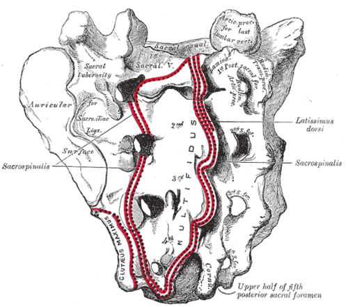
- Coccyx: small fused segments; attachment for the gluteus maximus, coccygeus and anococcygeal ligament
- Relations
The PSIS dimples overlie the SI joints at about S2. Anteriorly: rectum, sacral plexus, median sacral vessels.
Muscle attachments
- Gluteus maximus — origin
- Piriformis — origin
Joints
Ligament attachments
Mentioned in
Generated automatically from links in other entries.
Swipe sideways to see the whole diagram →

Clinical correlations
Caudal epidural block
- Mechanism / lesion
Local anesthetic injected through the sacral hiatus into the epidural space of the sacral canal.
- Classic presentation
Common in pediatric anesthesia and for some chronic pain procedures. The hiatus is palpated between the sacral cornua.
Coccydynia PANCE
- Mechanism / lesion
Pain at the coccyx from a fall onto the buttocks, childbirth, or prolonged sitting.
- Classic presentation
Localized pain over the coccyx, worse sitting (especially leaning back) and rising from sitting. Tenderness on palpation.
Pilonidal disease PANCE
- Mechanism / lesion
Hair and debris penetrating the skin of the natal cleft over the sacrococcygeal region, forming a sinus or abscess; common in young, hirsute men.
- Classic presentation
Painful, fluctuant swelling in the upper natal cleft, often with midline pits. Abscesses need incision and drainage.
Sacral insufficiency fracture
- Mechanism / lesion
Osteoporotic fracture of the sacrum in older adults (often after pelvic radiation or on steroids).
- Classic presentation
Low back, buttock or groin pain without major trauma; radiographs are often normal. MRI or CT shows the fracture.
High-yield PANCE pearls
- Dural sac ends at about S2; sacral hiatus → caudal epidural block.
- Coccydynia: fall or childbirth, pain with sitting.
- Pilonidal abscess: natal cleft, young hirsute men → incision and drainage.
Imaging anatomy
- X-ray
Pelvis AP and lateral sacrum/coccyx; sacral fractures are often hard to see.
- CT
Sacral fractures and SI joint detail.
- MRI
Insufficiency fractures (edema), sacroiliitis, sacral tumors (e.g. chordoma).
- Key views & landmarks
- Lateral sacrum/coccyx radiograph
- MRI or CT for suspected sacral fracture
Physical exam
- Surface landmarks
PSIS dimples at about S2; sacral hiatus palpable as a depression between the cornua at the upper natal cleft; coccyx tip in the natal cleft.
- Palpation
Palpate the coccyx externally (and per rectum for mobility) for tenderness. Inspect the natal cleft for pits and abscess.
Self-quiz
At approximately which level does the dural sac end in adults?
Correct answer: D
The spinal cord ends at about L1–L2, but the dural sac (and subarachnoid space) continues to about S2.
Show answer
D
The spinal cord ends at about L1–L2, but the dural sac (and subarachnoid space) continues to about S2.
Through which opening is local anesthetic introduced for a caudal epidural block?
Correct answer: B
The sacral hiatus, between the sacral cornua, leads into the sacral epidural space.
Show answer
B
The sacral hiatus, between the sacral cornua, leads into the sacral epidural space.
A 20-year-old man has a painful, red, fluctuant swelling in the upper natal cleft with small midline pits. What is the most likely diagnosis?
Correct answer: B
A fluctuant swelling with midline pits in the natal cleft is a pilonidal abscess; treat with incision and drainage.
Show answer
B
A fluctuant swelling with midline pits in the natal cleft is a pilonidal abscess; treat with incision and drainage.