Femoral triangle
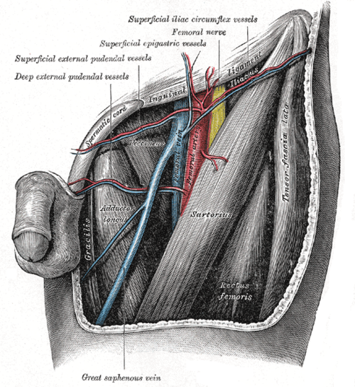
Triangular region of the upper anterior thigh containing the femoral nerve, artery, vein and canal. It is the site of the femoral pulse, arterial and venous access, and femoral hernias.
Core anatomy
- Location
Upper anterior thigh, just below the inguinal ligament.
- Boundaries
- superior (base)
- Inguinal ligament
- lateral
- Medial border of sartorius
- medial
- Medial border of adductor longus
- floor
- Iliopsoas (laterally) and pectineus (medially), with adductor longus
- roof
- Fascia lata with the saphenous opening (cribriform fascia)
- apex
- Where sartorius crosses adductor longus, continuing as the adductor canal
- Contents
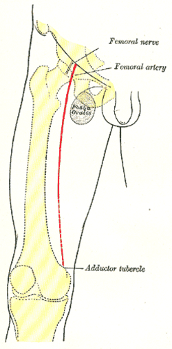
- Lateral to medial — NAVEL: Femoral nerve (outside the sheath), femoral artery, femoral vein, empty space/femoral canal, lymphatics (deep inguinal nodes, node of Cloquet)
- Femoral sheath: a fascial sleeve (from transversalis and iliac fascia) around the artery, vein and canal — not the nerve
- Deep femoral artery and vein; the termination of the great saphenous vein through the saphenous opening
- Femoral branch of the genitofemoral nerve
- Communications
Above: the femoral ring (the mouth of the femoral canal) opens into the abdomen — bounded by the inguinal ligament (anterior), pectineal ligament (posterior), lacunar ligament (medial) and femoral vein (lateral). Below: continuous with the adductor canal.
Mentioned in
Generated automatically from links in other entries.
Swipe sideways to see the whole diagram →


Clinical correlations
Femoral hernia PANCE
- Mechanism / lesion
Herniation through the femoral ring into the femoral canal, below the inguinal ligament and medial to the femoral vein; more common in women.
- Classic presentation
A small groin bulge below and lateral to the pubic tubercle; highest risk of incarceration and strangulation of groin hernias (narrow, rigid ring), so repair is recommended. See inguinal canal.
Femoral vascular access PANCE
- Mechanism / lesion
Arterial puncture for angiography/PCI; central venous access into the femoral vein.
- Classic presentation
Palpate the arterial pulse below the mid-inguinal point; the vein is just medial. Puncture below the inguinal ligament — a high puncture can cause retroperitoneal hemorrhage.
Inguinal lymphadenopathy
- Mechanism / lesion
Drainage of the lower limb, perineum, lower abdominal wall and anal canal below the pectinate line (see pelvic and inguinal lymph drainage).
- Classic presentation
Enlarged nodes in the triangle; examine the whole drainage area.
High-yield PANCE pearls
- NAVEL lateral → medial: Nerve, Artery, Vein, Empty space, Lymphatics.
- Femoral hernia: women, below the inguinal ligament, medial to the femoral vein, highest strangulation risk.
- Femoral venous line: vein is medial to the pulse.
- The femoral nerve lies outside the femoral sheath.
Imaging anatomy
- CT
Femoral hernias with bowel obstruction; hematomas after catheterization.
- Ultrasound
Guides femoral arterial and venous access and nerve blocks; distinguishes hernia, lymph node, pseudoaneurysm and saphena varix.
Physical exam
- Surface landmarks
Inguinal ligament from the ASIS to the pubic tubercle; the femoral pulse just below its midpoint (mid-inguinal point).
- Palpation
Femoral pulse; lymph nodes; groin masses (a femoral hernia lies below and lateral to the pubic tubercle; an inguinal hernia above and medial). A saphena varix has a cough thrill and disappears on lying down.
- Strength / function testing
Cough impulse over any groin mass.
Self-quiz
Which structure lies immediately medial to the femoral vein in the femoral triangle?
Correct answer: C
Lateral to medial: nerve, artery, vein, femoral canal (empty space), lymphatics. Femoral hernias pass through the canal, just medial to the vein.
Show answer
C
Lateral to medial: nerve, artery, vein, femoral canal (empty space), lymphatics. Femoral hernias pass through the canal, just medial to the vein.
A 70-year-old woman has vomiting and a tender, irreducible lump in the groin below the inguinal ligament and lateral to the pubic tubercle. What is the most likely diagnosis?
Correct answer: C
A lump below the inguinal ligament and inferolateral to the pubic tubercle in an older woman with bowel obstruction is an incarcerated femoral hernia — the groin hernia most prone to strangulation, needing urgent surgery.
Show answer
C
A lump below the inguinal ligament and inferolateral to the pubic tubercle in an older woman with bowel obstruction is an incarcerated femoral hernia — the groin hernia most prone to strangulation, needing urgent surgery.
Which muscle forms the medial boundary of the femoral triangle?
Correct answer: A
The femoral triangle is bounded by the inguinal ligament (base), the medial border of sartorius (lateral) and the medial border of adductor longus (medial). Iliopsoas, pectineus and adductor longus form its floor.
Show answer
A
The femoral triangle is bounded by the inguinal ligament (base), the medial border of sartorius (lateral) and the medial border of adductor longus (medial). Iliopsoas, pectineus and adductor longus form its floor.
Which structure forms the medial boundary of the femoral ring, the opening of the femoral canal?
Correct answer: C
The femoral ring is bounded by the inguinal ligament (anterior), the pectineal ligament (posterior), the lacunar ligament (medial) and the femoral vein (lateral). The rigid, narrow ring explains why femoral hernias strangulate so often.
Show answer
C
The femoral ring is bounded by the inguinal ligament (anterior), the pectineal ligament (posterior), the lacunar ligament (medial) and the femoral vein (lateral). The rigid, narrow ring explains why femoral hernias strangulate so often.
Where should the needle be placed to cannulate the femoral vein?
Correct answer: D
From lateral to medial the triangle contains the Nerve, Artery, Vein, Empty space (femoral canal) and Lymphatics (NAVEL). The vein lies just medial to the arterial pulse, and puncture should be below the inguinal ligament.
Show answer
D
From lateral to medial the triangle contains the Nerve, Artery, Vein, Empty space (femoral canal) and Lymphatics (NAVEL). The vein lies just medial to the arterial pulse, and puncture should be below the inguinal ligament.
Flashcards
See also
- Femoral artery
- Femoral nerve
- Great saphenous vein
- Inguinal canal
- Groin hernias (comparison table)