Gallbladder and biliary tree
Pear-shaped sac under the liver that stores and concentrates bile, with the extrahepatic bile ducts. Gallstone disease (biliary colic, cholecystitis, choledocholithiasis, cholangitis) is one of the most tested topics.
Core anatomy
- Location
In the gallbladder fossa on the visceral surface of the liver. The fundus projects at the tip of the right 9th costal cartilage, where the lateral border of the right rectus abdominis meets the costal margin.
- Parts
- Fundus, body, neck (with Hartmann pouch, a common site of impacted stones)
- Cystic duct (with spiral folds, the spiral valve of Heister) joins the common hepatic duct to form the common bile duct (CBD)
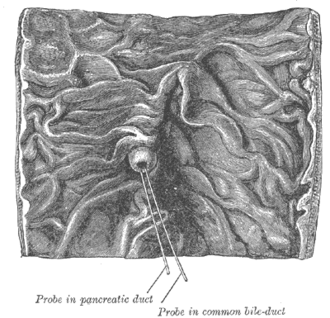
- The CBD descends in the hepatoduodenal ligament, passes posterior to the 1st part of the duodenum, runs through or behind the head of the pancreas, and joins the main pancreatic duct to form the hepatopancreatic ampulla (of Vater), opening at the major duodenal papilla in the 2nd part of the duodenum. The sphincter of Oddi controls flow
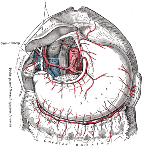
- Cystohepatic triangle (of Calot): cystic duct, common hepatic duct, and the inferior surface of the liver. It contains the cystic artery (usually from the right hepatic artery) and must be clearly identified in cholecystectomy (the "critical view of safety")
- Relations
The fundus contacts the transverse colon; the body contacts the 1st and 2nd parts of the duodenum (the site of cholecystoenteric fistula in gallstone ileus).
- Arterial supply
Cystic artery, usually from the right hepatic artery.
- Venous drainage
Cystic veins and small veins directly into the liver.
- Innervation
Celiac plexus (sympathetic, visceral pain in the epigastrium and RUQ), vagus, and the right phrenic nerve (referred pain to the right shoulder or scapula).
Swipe sideways to see the whole diagram →


Clinical correlations
Biliary colic and acute cholecystitis PANCE
- Mechanism / lesion
Biliary colic: transient obstruction of the cystic duct by a stone. Acute cholecystitis: persistent obstruction with gallbladder inflammation. Risk factors ("5 F's"): female, fertile, forty, fat (obesity), fair; also rapid weight loss, pregnancy, estrogen and hemolysis (pigment stones).
- Classic presentation
Colic: postprandial (fatty meal) RUQ/epigastric pain lasting less than about 6 hours, radiating to the right scapula. Cholecystitis: longer pain, fever, leukocytosis, Murphy sign.
Choledocholithiasis and ascending cholangitis PANCE
- Mechanism / lesion
Stones in the common bile duct obstruct bile flow; superimposed infection causes cholangitis.
- Classic presentation
Jaundice, elevated bilirubin and alkaline phosphatase. Cholangitis: Charcot triad (fever, RUQ pain, jaundice); Reynolds pentad adds hypotension and altered mental status. Needs antibiotics and urgent biliary drainage (ERCP).
Gallstone pancreatitis and gallstone ileus PANCE
- Mechanism / lesion
A stone impacted at the ampulla blocks the pancreatic duct (pancreatitis). A large stone eroding through a cholecystoenteric fistula can obstruct the terminal ileum (gallstone ileus).
- Classic presentation
Pancreatitis: epigastric pain radiating to the back with elevated lipase. Gallstone ileus: small bowel obstruction with pneumobilia and an ectopic stone (Rigler triad) in an older woman.
Courvoisier sign PANCE
- Mechanism / lesion
Gradual distal CBD obstruction by malignancy (pancreatic head cancer, cholangiocarcinoma) distends a compliant gallbladder. Chronic gallstone disease causes a fibrotic, non-distensible gallbladder.
- Classic presentation
A palpable, non-tender gallbladder with painless jaundice suggests malignancy rather than stones.
High-yield PANCE pearls
- Ultrasound is first-line for RUQ pain / suspected cholecystitis; HIDA if ultrasound is equivocal (non-filling gallbladder = cholecystitis).
- Charcot triad: fever, RUQ pain, jaundice → cholangitis → ERCP. Reynolds pentad adds hypotension + confusion.
- Murphy sign: inspiratory arrest with RUQ palpation.
- Courvoisier: painless jaundice + palpable non-tender gallbladder → pancreatic head cancer.
- Calot triangle: cystic duct, common hepatic duct, liver edge; contains the cystic artery.
Imaging anatomy
- X-ray
Most gallstones are not radiopaque (only about 10–15% are visible). Pneumobilia with gallstone ileus. Porcelain gallbladder (calcified wall). verify
- CT
Less sensitive than ultrasound for gallstones. Useful for complications (perforation, emphysematous cholecystitis, abscess), malignancy, and pancreatitis.
- MRI
MRCP noninvasively shows the biliary tree and CBD stones before choosing ERCP.
- Ultrasound
First-line. Gallstones are echogenic with posterior acoustic shadowing and move with position. Acute cholecystitis: stones plus wall thickening (more than about 3 mm), pericholecystic fluid, gallbladder distension and a sonographic Murphy sign. A dilated CBD (more than about 6 mm, higher after cholecystectomy or with age) suggests obstruction. verify
- Key views & landmarks
- RUQ ultrasound
- HIDA (cholescintigraphy) if ultrasound equivocal
- MRCP / ERCP (therapeutic) for CBD stones
Physical exam
- Surface landmarks
The gallbladder point: tip of the right 9th costal cartilage, at the lateral border of rectus abdominis (linea semilunaris).
- Palpation
Palpate at the gallbladder point. A palpable gallbladder with jaundice suggests Courvoisier sign.
Murphy sign
- Technique
Place fingers under the right costal margin at the gallbladder point and ask the patient to breathe in deeply.
- Positive test
The patient stops inspiring abruptly (catches the breath) because of pain as the inflamed gallbladder descends onto the fingers.
- Significance
Acute cholecystitis. Less sensitive in older adults.
Self-quiz
A 42-year-old woman has 12 hours of constant RUQ pain after a fatty meal, fever of 38.5 °C, and leukocytosis. During palpation below the right costal margin she stops inhaling abruptly. What is the best initial imaging?
Correct answer: B
Suspected acute cholecystitis (positive Murphy sign, fever) is evaluated first with RUQ ultrasound. HIDA is used if ultrasound is equivocal. ERCP is for CBD stones.
Show answer
B
Suspected acute cholecystitis (positive Murphy sign, fever) is evaluated first with RUQ ultrasound. HIDA is used if ultrasound is equivocal. ERCP is for CBD stones.
A 70-year-old woman has fever, RUQ pain and jaundice, and she becomes hypotensive and confused. What is the definitive treatment after resuscitation and antibiotics?
Correct answer: B
Reynolds pentad indicates severe ascending cholangitis. After antibiotics and fluids, the patient needs urgent biliary drainage (ERCP).
Show answer
B
Reynolds pentad indicates severe ascending cholangitis. After antibiotics and fluids, the patient needs urgent biliary drainage (ERCP).
A 66-year-old man has painless jaundice, weight loss, and a palpable, non-tender gallbladder. What is the most likely diagnosis?
Correct answer: C
Courvoisier sign (painless jaundice + palpable non-tender gallbladder) suggests malignant distal CBD obstruction, most commonly pancreatic head carcinoma.
Show answer
C
Courvoisier sign (painless jaundice + palpable non-tender gallbladder) suggests malignant distal CBD obstruction, most commonly pancreatic head carcinoma.
Which structure is found within the cystohepatic triangle (of Calot)?
Correct answer: B
The triangle (cystic duct, common hepatic duct, inferior liver surface) contains the cystic artery, which is ligated during cholecystectomy.
Show answer
B
The triangle (cystic duct, common hepatic duct, inferior liver surface) contains the cystic artery, which is ligated during cholecystectomy.
An 80-year-old woman has small bowel obstruction. CT shows air in the biliary tree and a large gallstone in the bowel. Where is the stone most likely lodged?
Correct answer: D
In gallstone ileus, a large stone erodes through a cholecystoenteric fistula (gallbladder body to the duodenum) and obstructs the terminal ileum, the narrowest part. Pneumobilia, obstruction and an ectopic stone form Rigler triad.
Show answer
D
In gallstone ileus, a large stone erodes through a cholecystoenteric fistula (gallbladder body to the duodenum) and obstructs the terminal ileum, the narrowest part. Pneumobilia, obstruction and an ectopic stone form Rigler triad.
Flashcards
See also
- Liver
- Pancreas
- Duodenum
- Foregut, midgut and hindgut (comparison table)