Uterus
Thick-walled muscular organ between the bladder and rectum in which the embryo implants and develops. Fibroids, endometrial cancer, cervical cancer and postpartum hemorrhage are its key clinical problems.
Core anatomy
- Location
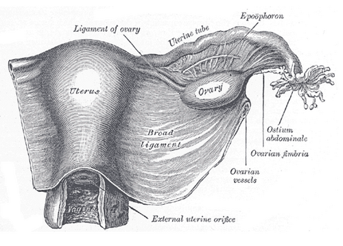
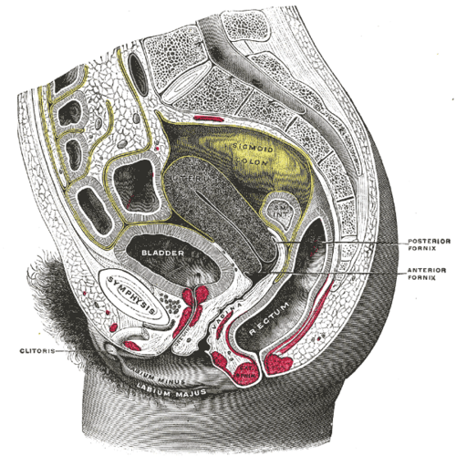
In the lesser pelvis, between the urinary bladder (anteriorly) and the rectum (posteriorly), with the broad ligament extending from each side to the pelvic wall. Usually anteverted (the cervix angled forward on the vagina) and anteflexed (the body bent forward on the cervix), so it rests on the bladder.
- Parts
- Fundus: above the openings of the uterine tubes
- Body (corpus): the main part; the uterine cavity is triangular in coronal section
- Isthmus: the narrow part between the body and cervix; becomes the lower uterine segment in late pregnancy (site of the low transverse cesarean incision)
- Cervix: supravaginal part and vaginal part (portio) with the external os; cervical canal from the internal to the external os. The transformation zone (at the squamocolumnar junction) is where cervical dysplasia arises
- Wall: endometrium (functional and basal layers), myometrium, perimetrium (peritoneum)
- Supports: the pelvic diaphragm and the endopelvic fascial ligaments — cardinal (transverse cervical) ligaments (carrying the uterine vessels), uterosacral and pubocervical ligaments. The round ligament and broad ligament give little support
- Relations
Anteriorly the vesicouterine pouch; posteriorly the rectouterine pouch. The ureter passes under the uterine artery about 1–2 cm lateral to the supravaginal cervix ("water under the bridge").
- Arterial supply
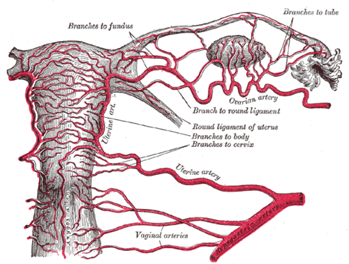
Uterine arteries (anterior division of the internal iliac artery), anastomosing with the ovarian arteries at the uterotubal junction.
- Venous drainage
Uterine venous plexus in the broad ligament → uterine veins → internal iliac veins.
- Lymphatic drainage
Fundus and upper body → lumbar (para-aortic) nodes along the ovarian vessels; some along the round ligament to the superficial inguinal nodes. Body → external iliac nodes. Cervix → internal and external iliac (obturator) and sacral nodes. See pelvic and inguinal lymph drainage.
- Innervation
Uterovaginal plexus (from the inferior hypogastric plexus). Pain from the uterine body (above the pelvic pain line) travels with sympathetic fibers to about T10–L1 (first-stage labor pain); pain from the cervix travels with the pelvic splanchnic nerves (S2–S4). See pelvic autonomic nerves.
Mentioned in
Generated automatically from links in other entries.
Swipe sideways to see the whole diagram →



Clinical correlations
Leiomyoma (fibroids) PANCE
- Mechanism / lesion
Benign, estrogen-sensitive smooth muscle tumors of the myometrium; the most common pelvic tumor in women, more common in Black women; shrink after menopause.
- Classic presentation
Often asymptomatic. Heavy menstrual bleeding, pelvic pressure, urinary frequency, constipation, infertility. Exam: enlarged, irregular, firm uterus. Diagnosed by pelvic ultrasound.
Endometrial cancer PANCE
- Mechanism / lesion
Most commonly endometrioid adenocarcinoma driven by unopposed estrogen — obesity, nulliparity, early menarche/late menopause, tamoxifen, chronic anovulation (PCOS), Lynch syndrome.
- Classic presentation
Postmenopausal bleeding (any bleeding after menopause must be evaluated). Evaluation: transvaginal ultrasound (endometrial thickness ≤ 4 mm is reassuring) and/or endometrial biopsy.
Cervical cancer and screening PANCE
- Mechanism / lesion
Persistent high-risk HPV (types 16 and 18) infection of the transformation zone → dysplasia (CIN) → squamous cell carcinoma (most) or adenocarcinoma.
- Classic presentation
Early disease is asymptomatic; later postcoital bleeding, discharge, pelvic pain. Screening (USPSTF 2018): cytology every 3 years at 21–29; at 30–65, cytology every 3 years, primary hrHPV testing every 5 years, or co-testing every 5 years. HPV vaccination prevents most cases.
Postpartum hemorrhage PANCE
- Mechanism / lesion
Uterine atony is the most common cause (the "4 Ts": tone, trauma, tissue, thrombin). Contraction of the myometrium normally compresses the spiral arteries ("living ligatures").
- Classic presentation
Excessive bleeding after delivery with a soft, boggy uterus. Management: bimanual uterine massage, oxytocin, then other uterotonics (methylergonovine — avoid in hypertension; carboprost — avoid in asthma), tranexamic acid, then procedures (balloon tamponade, uterine artery embolization or ligation, hysterectomy).
Adenomyosis
- Mechanism / lesion
Endometrial glands and stroma within the myometrium.
- Classic presentation
Dysmenorrhea and heavy menstrual bleeding in parous women in their 40s; symmetrically enlarged, boggy, tender uterus.
High-yield PANCE pearls
- Postmenopausal bleeding → TVUS and/or endometrial biopsy to exclude endometrial cancer.
- Enlarged irregular uterus = fibroids; symmetric boggy tender uterus = adenomyosis.
- Most common cause of postpartum hemorrhage = uterine atony → massage + oxytocin.
- Cervical cancer = HPV 16/18; screening starts at 21.
- Fundal height: at the umbilicus at ~20 weeks; in cm ≈ weeks of gestation from about 20–36 weeks.
Imaging anatomy
- X-ray
Hysterosalpingography (HSG) shows the uterine cavity (congenital anomalies, synechiae) and tubal patency.
- MRI
Maps fibroids before surgery or embolization, and characterizes adenomyosis; stages endometrial and cervical cancer.
- Ultrasound
Transvaginal ultrasound is first-line: uterine size, fibroids, endometrial thickness, adenomyosis, early pregnancy. Saline infusion sonohysterography shows polyps and submucosal fibroids.
- Key views & landmarks
- Transvaginal ultrasound (sagittal and transverse)
- MRI pelvis (T2) for fibroid mapping and cancer staging
Physical exam
- Surface landmarks
The non-pregnant uterus is not palpable abdominally. In pregnancy the fundus reaches the pubic symphysis at about 12 weeks and the umbilicus at about 20 weeks; fundal height in cm approximates gestational age from about 20 to 36 weeks.
- Palpation
Bimanual exam: size (in "weeks" equivalent), shape (regular or irregular), consistency, mobility, position (anteverted or retroverted) and tenderness. Cervical motion tenderness suggests PID or ectopic pregnancy.
- Strength / function testing
Speculum exam to inspect the cervix and take cervical cytology/HPV samples.
Self-quiz
A 58-year-old woman, 6 years postmenopausal, has had two episodes of light vaginal bleeding. BMI is 38. What is the most appropriate next step?
Correct answer: C
Postmenopausal bleeding must be evaluated for endometrial cancer, especially with obesity (peripheral estrogen from adipose tissue). Transvaginal ultrasound (endometrial thickness) and/or endometrial biopsy are the initial tests.
Show answer
C
Postmenopausal bleeding must be evaluated for endometrial cancer, especially with obesity (peripheral estrogen from adipose tissue). Transvaginal ultrasound (endometrial thickness) and/or endometrial biopsy are the initial tests.
Twenty minutes after a vaginal delivery, a woman has heavy bleeding. The uterus is soft and boggy above the umbilicus. What is the most appropriate first step?
Correct answer: B
A boggy uterus indicates uterine atony, the most common cause of postpartum hemorrhage. First-line treatment is bimanual massage and oxytocin; other uterotonics and procedures follow if bleeding continues.
Show answer
B
A boggy uterus indicates uterine atony, the most common cause of postpartum hemorrhage. First-line treatment is bimanual massage and oxytocin; other uterotonics and procedures follow if bleeding continues.
Lymph from the uterine fundus drains mainly to which nodes?
Correct answer: C
The fundus and upper body drain along the ovarian vessels to the lumbar (para-aortic) nodes. Some fundal lymph follows the round ligament to the superficial inguinal nodes, and the cervix drains to the iliac and sacral nodes.
Show answer
C
The fundus and upper body drain along the ovarian vessels to the lumbar (para-aortic) nodes. Some fundal lymph follows the round ligament to the superficial inguinal nodes, and the cervix drains to the iliac and sacral nodes.
A 44-year-old woman who has had three children has worsening dysmenorrhea and heavy menstrual bleeding. The uterus is symmetrically enlarged, boggy and tender. What is the most likely diagnosis?
Correct answer: B
Adenomyosis (endometrial glands and stroma within the myometrium) causes dysmenorrhea and heavy bleeding in parous women in their 40s, with a symmetrically enlarged, boggy, tender uterus. Fibroids give an enlarged, irregular, firm uterus.
Show answer
B
Adenomyosis (endometrial glands and stroma within the myometrium) causes dysmenorrhea and heavy bleeding in parous women in their 40s, with a symmetrically enlarged, boggy, tender uterus. Fibroids give an enlarged, irregular, firm uterus.
At about how many weeks of gestation does the uterine fundus reach the level of the umbilicus?
Correct answer: C
The fundus reaches the umbilicus at about 20 weeks. From about 20 to 36 weeks, fundal height in centimeters roughly equals the weeks of gestation.
Show answer
C
The fundus reaches the umbilicus at about 20 weeks. From about 20 to 36 weeks, fundal height in centimeters roughly equals the weeks of gestation.
Flashcards
See also
- Uterine artery
- Broad ligament
- Uterine tubes
- Ovary
- Vagina
- Lymph drainage of the pelvis and perineum (comparison table)
- Collapse in early pregnancy (clinical case)