Inguinal canal
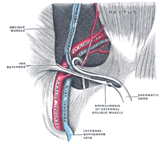
Oblique passage through the lower anterior abdominal wall carrying the spermatic cord or round ligament. It's the site of indirect and direct inguinal hernias, the most common hernias overall.
Core anatomy
- Location
Above and parallel to the medial half of the inguinal ligament, about 4 cm long, running inferomedially from the deep ring to the superficial ring.
- Boundaries
- deep ring
- Opening in the transversalis fascia, just above the midpoint of the inguinal ligament and lateral to the inferior epigastric vessels
- superficial ring
- Opening in the external oblique aponeurosis, just superolateral to the pubic tubercle
- anterior wall
- External oblique aponeurosis (reinforced laterally by internal oblique)
- posterior wall
- Transversalis fascia (reinforced medially by the conjoint tendon)
- roof
- Arching fibers of internal oblique and transversus abdominis
- floor
- Inguinal ligament (with the lacunar ligament medially)
- Contents
- Males: Spermatic cord
- Females: round ligament of the uterus
- Both: ilioinguinal nerve (enters the canal partway along, not through the deep ring, and exits the superficial ring); genital branch of the genitofemoral nerve (within the cord in males)
- Communications
From the extraperitoneal space (deep ring) to the scrotum or labium majus (superficial ring).
- Notes
Mnemonic for the walls, MALT from superior clockwise: roof Muscles (internal oblique, transversus), anterior Aponeurosis (external oblique), floor Ligament (inguinal), posterior Transversalis fascia. The canal's obliquity and the muscle "shutter" mechanism normally resist herniation.
Found in
Mentioned in
- Anterior abdominal wall
- Ductus deferens and seminal glands
- Femoral triangle
- Lumbar plexus
- Spermatic cord
- Testis and epididymis
Generated automatically from links in other entries.
Swipe sideways to see the whole diagram →

Clinical correlations
Indirect inguinal hernia PANCE
- Mechanism / lesion
Viscera enter the deep ring lateral to the inferior epigastric vessels and follow the canal, often through a patent processus vaginalis (congenital). The most common hernia in both sexes and in children. Can descend into the scrotum.
- Classic presentation
A groin bulge that may extend into the scrotum, enlarging with coughing or straining. In infants, an intermittent groin or scrotal swelling while crying.
Direct inguinal hernia PANCE
- Mechanism / lesion
Protrusion through a weak posterior wall (transversalis fascia) in the inguinal triangle, medial to the inferior epigastric vessels. Acquired, usually in older men.
- Classic presentation
A bulge in the medial groin that rarely enters the scrotum. Lower strangulation risk than indirect or femoral hernias.
Femoral hernia (for contrast) PANCE
- Mechanism / lesion
Passes below the inguinal ligament through the femoral ring into the femoral canal, medial to the femoral vein. More common in women.
- Classic presentation
A small bulge below and lateral to the pubic tubercle. Highest risk of incarceration and strangulation of groin hernias, so repair is recommended.
High-yield PANCE pearls
- Indirect = through the deep ring, lateral to the inferior epigastrics, patent processus vaginalis, most common (including in women and children).
- Direct = Hesselbach triangle, medial to the inferior epigastrics, older men.
- Femoral = below the inguinal ligament, women, highest strangulation risk.
- Canal walls: MALT (Muscles, Aponeurosis, Ligament, Transversalis fascia).
- Incarcerated/strangulated hernia (tender, irreducible, erythema, obstruction) → emergency surgery.
Imaging anatomy
- CT
Shows hernia contents and complications (bowel obstruction, strangulation). Helpful in obese patients or equivocal exams.
- MRI
Occult hernias and athletic groin pain ("sports hernia" or athletic pubalgia) when ultrasound is inconclusive.
- Ultrasound
Dynamic ultrasound with Valsalva is first-line imaging for an equivocal groin bulge: it locates the sac relative to the inferior epigastric vessels (indirect vs direct) and distinguishes a femoral hernia (below the ligament, medial to the femoral vein).
- Key views & landmarks
- Dynamic groin ultrasound at rest and with Valsalva
Physical exam
- Surface landmarks
ASIS and pubic tubercle mark the ends of the inguinal ligament. The deep ring lies about 1.5 cm above the midpoint of the inguinal ligament. The superficial ring lies above and lateral to the pubic tubercle.
- Palpation
Examine standing. Inspect both groins while the patient coughs. In males, invaginate the loose scrotal skin with a finger up toward the superficial ring and ask the patient to cough or bear down; feel for an impulse. Examine for femoral hernias below the inguinal ligament.
- Strength / function testing
Attempt gentle reduction if not tender. The traditional teaching that an impulse at the fingertip means indirect and at the finger pad means direct is unreliable on exam; the distinction is made surgically or on imaging. verify
Self-quiz
A 6-month-old boy has an intermittent swelling in the right groin extending into the scrotum that appears when he cries. What is the underlying defect?
Correct answer: B
An indirect inguinal hernia in infants results from a patent processus vaginalis, which lets bowel follow the path of testicular descent through the deep ring.
Show answer
B
An indirect inguinal hernia in infants results from a patent processus vaginalis, which lets bowel follow the path of testicular descent through the deep ring.
Which structure forms the posterior wall of the inguinal canal?
Correct answer: C
The transversalis fascia (reinforced medially by the conjoint tendon) forms the posterior wall. Weakness of it in the inguinal triangle produces direct hernias.
Show answer
C
The transversalis fascia (reinforced medially by the conjoint tendon) forms the posterior wall. Weakness of it in the inguinal triangle produces direct hernias.
A 70-year-old woman has a small, tender, irreducible bulge just below the inguinal ligament, lateral to the pubic tubercle, with vomiting. What is the most likely diagnosis?
Correct answer: C
A bulge below the inguinal ligament in a woman is a femoral hernia, which has the highest risk of incarceration and strangulation. This one needs emergency repair.
Show answer
C
A bulge below the inguinal ligament in a woman is a femoral hernia, which has the highest risk of incarceration and strangulation. This one needs emergency repair.
Which nerve travels through the inguinal canal but does NOT pass through the deep inguinal ring?
Correct answer: B
The ilioinguinal nerve enters the canal from its lateral wall (not through the deep ring) and exits the superficial ring. It can be injured in open hernia repair, causing groin and scrotal/labial numbness or pain.
Show answer
B
The ilioinguinal nerve enters the canal from its lateral wall (not through the deep ring) and exits the superficial ring. It can be injured in open hernia repair, causing groin and scrotal/labial numbness or pain.
The superficial inguinal ring is an opening in which layer?
Correct answer: A
The superficial ring is an opening in the external oblique aponeurosis, just superolateral to the pubic tubercle. The deep ring is an opening in the transversalis fascia, lateral to the inferior epigastric vessels.
Show answer
A
The superficial ring is an opening in the external oblique aponeurosis, just superolateral to the pubic tubercle. The deep ring is an opening in the transversalis fascia, lateral to the inferior epigastric vessels.