Cerebellum
Coordinates movement, balance and eye movements. Hemisphere lesions cause ipsilateral limb ataxia, vermis lesions cause truncal and gait ataxia, and cerebellar strokes can swell and compress the brainstem.
Core anatomy
- Location
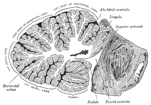
In the posterior cranial fossa under the tentorium cerebelli, behind the pons, medulla oblongata and the fourth ventricle.
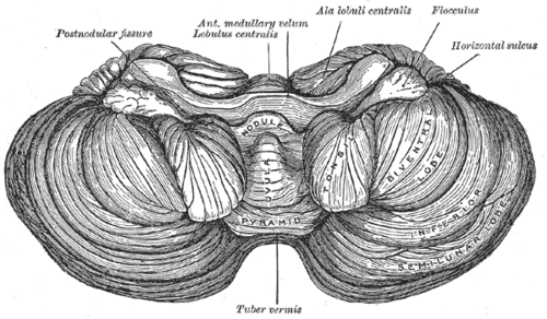
- Parts
- Vermis (midline) — trunk, posture, gait (spinocerebellum); anterior vermis damaged by alcohol
- Hemispheres (lateral) — planning and coordination of ipsilateral limb movements (cerebrocerebellum)
- Flocculonodular lobe — balance and eye movements (vestibulocerebellum)
- Deep nuclei (lateral to medial): dentate, emboliform, globose, fastigial ("Don't Eat Greasy Foods")
- Peduncles: superior (mainly output — dentate → contralateral red nucleus and thalamus (VL) → motor cortex); middle (input from the contralateral pontine nuclei — the largest); inferior (input from the spinal cord, vestibular nuclei and inferior olive)
- Cortex: Purkinje cells — the sole output of the cortex, inhibitory (GABA) onto the deep nuclei; tonsils above the foramen magnum
- Relations
The fourth ventricle lies in front — cerebellar swelling obstructs it, causing hydrocephalus and brainstem compression.
- Arterial supply
Superior cerebellar artery (SCA), anterior inferior cerebellar artery (AICA) and posterior inferior cerebellar artery (PICA) — see vertebrobasilar system.
- Venous drainage
Cerebellar veins → straight, transverse and petrosal sinuses.
- Notes
Because output crosses to the opposite thalamus and cortex, whose corticospinal fibers cross back, each cerebellar hemisphere controls the same side of the body (lesions → ipsilateral signs).
Mentioned in
Generated automatically from links in other entries.


Clinical correlations
Cerebellar stroke or hemorrhage PANCE
- Mechanism / lesion
Infarction (PICA, SCA, AICA) or hypertensive hemorrhage.
- Classic presentation
Sudden vertigo, vomiting, headache, inability to stand or walk, ipsilateral limb ataxia, nystagmus, dysarthria. Can mimic peripheral vertigo (use HINTS). Swelling over 2–4 days can compress the fourth ventricle and brainstem → declining consciousness → emergency suboccipital decompression or ventricular drain.
Alcohol-related cerebellar degeneration PANCE
- Mechanism / lesion
Chronic alcohol use (± thiamine deficiency) → atrophy of the anterior superior vermis.
- Classic presentation
Wide-based, unsteady gait and truncal ataxia with relatively normal arm coordination.
Posterior fossa tumors in children PANCE
- Mechanism / lesion
Medulloblastoma (vermis, most common malignant brain tumor of childhood), pilocytic astrocytoma (hemisphere, cystic, benign), ependymoma (fourth ventricle).
- Classic presentation
Morning headache and vomiting (hydrocephalus), truncal ataxia (vermis) or limb ataxia (hemisphere), papilledema.
High-yield PANCE pearls
- Cerebellar lesions → ipsilateral signs.
- Vermis → truncal/gait ataxia; hemisphere → limb ataxia, intention tremor, dysdiadochokinesia.
- Vertigo + vomiting + can't walk → rule out cerebellar stroke (MRI, not CT).
- Alcohol → anterior vermis → wide-based gait.
- Romberg is negative in pure cerebellar ataxia (unsteady with eyes open too); positive in dorsal column/sensory ataxia.
- Child with morning headaches, vomiting and ataxia → medulloblastoma (vermis).
Imaging anatomy
- CT
CT detects hemorrhage and hydrocephalus but can miss small posterior fossa infarcts.
- MRI
MRI with DWI is best for posterior fossa strokes and tumors.
Physical exam
- Strength / function testing
Finger-to-nose (dysmetria, intention tremor), heel-to-shin, rapid alternating movements (dysdiadochokinesia), rebound; gait and tandem walking (truncal ataxia); speech (scanning dysarthria); nystagmus (gaze-evoked). Signs mnemonic DANISH: Dysdiadochokinesia, Ataxia, Nystagmus, Intention tremor, Slurred speech, Hypotonia.
Romberg test
- Technique
Patient stands with feet together, arms at the sides; observe with the eyes open, then closed for about 30 seconds.
- Positive test
Loses balance only when the eyes are closed.
- Significance
Proprioceptive (dorsal column or peripheral sensory) ataxia — e.g. B12 deficiency, tabes dorsalis, neuropathy. Patients with cerebellar ataxia are unsteady even with the eyes open, so Romberg is not a test of the cerebellum.
Self-quiz
A 58-year-old has sudden vertigo and vomiting. He cannot walk, and finger-to-nose testing shows marked overshooting with the right hand. Which area is affected?
Correct answer: B
Right limb ataxia points to the right cerebellar hemisphere — cerebellar lesions cause ipsilateral signs. Sudden onset with vomiting and inability to walk suggests a cerebellar stroke: obtain MRI and watch for swelling.
Show answer
B
Right limb ataxia points to the right cerebellar hemisphere — cerebellar lesions cause ipsilateral signs. Sudden onset with vomiting and inability to walk suggests a cerebellar stroke: obtain MRI and watch for swelling.
A 55-year-old with chronic alcohol use has a wide-based unsteady gait. Finger-to-nose testing is normal. Which part of the cerebellum is most affected?
Correct answer: C
Alcohol preferentially damages the anterior (superior) vermis, which controls the trunk and gait, so gait ataxia occurs with relatively normal limb coordination.
Show answer
C
Alcohol preferentially damages the anterior (superior) vermis, which controls the trunk and gait, so gait ataxia occurs with relatively normal limb coordination.
How does the Romberg test typically behave in a patient with pure cerebellar ataxia?
Correct answer: B
A positive Romberg (steady with eyes open, falls with them closed) indicates sensory (proprioceptive) ataxia. In cerebellar ataxia the patient is unsteady even with the eyes open, so the test is classically negative.
Show answer
B
A positive Romberg (steady with eyes open, falls with them closed) indicates sensory (proprioceptive) ataxia. In cerebellar ataxia the patient is unsteady even with the eyes open, so the test is classically negative.
A 6-year-old has morning headaches, vomiting and a wide-based, unsteady gait. MRI shows a midline posterior fossa mass in the vermis. What is the most likely diagnosis?
Correct answer: D
Medulloblastoma arises in the vermis and is the most common malignant brain tumor of childhood; it causes truncal ataxia and hydrocephalus. Pilocytic astrocytomas are typically cystic tumors of the hemisphere.
Show answer
D
Medulloblastoma arises in the vermis and is the most common malignant brain tumor of childhood; it causes truncal ataxia and hydrocephalus. Pilocytic astrocytomas are typically cystic tumors of the hemisphere.
Which is the largest cerebellar peduncle, carrying input from the contralateral pontine nuclei?
Correct answer: C
The middle cerebellar peduncle is the largest and carries pontocerebellar fibers from the contralateral pontine nuclei. The superior peduncle is mainly output; the inferior peduncle carries spinal, vestibular and olivary input.
Show answer
C
The middle cerebellar peduncle is the largest and carries pontocerebellar fibers from the contralateral pontine nuclei. The superior peduncle is mainly output; the inferior peduncle carries spinal, vestibular and olivary input.