Ankle joint
Hinge joint in which the talus sits in a mortise formed by the tibia and fibula. Sprains of the lateral ligaments and malleolar fractures are extremely common; the Ottawa rules decide who needs an X-ray.
Core anatomy
- Location
Between the leg and the foot; the malleoli are palpable on each side.
- Classification
Synovial hinge joint
- Articulating surfaces
The distal tibia (plafond and medial malleolus), the fibula (lateral malleolus) and the trochlea of the talus (bones of the foot)
- Capsule
Thin anteriorly and posteriorly, reinforced on each side by the collateral ligaments.
- Ligaments
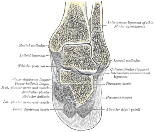
- Medial (deltoid) ligament: strong, fan-shaped, from the medial malleolus to the navicular, talus and calcaneus; resists eversion
- Lateral ankle ligaments: anterior talofibular (ATFL), calcaneofibular (CFL) and posterior talofibular (PTFL); resist inversion
- Distal tibiofibular syndesmosis: anterior and posterior inferior tibiofibular ligaments and the interosseous ligament, holding the mortise together
- Movements
- Dorsiflexion (tibialis anterior, extensor digitorum and hallucis longus, fibularis tertius) — about 20°
- Plantarflexion (triceps surae and calcaneal tendon, tibialis posterior, flexor hallucis and digitorum longus, fibularis longus and brevis) — about 50°
- Inversion and eversion occur mainly at the subtalar and transverse tarsal joints, not the ankle
- Stability
The talus is wider anteriorly, so the joint is most stable in dorsiflexion and least stable in plantarflexion — which is why most sprains happen with the foot plantarflexed and inverted.
- Innervation
Deep fibular and tibial nerves (branches to the joint).
- Blood supply
Malleolar branches of the anterior tibial, posterior tibial and fibular arteries.
- Relations
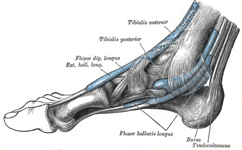
Behind the medial malleolus (tarsal tunnel, front to back — "Tom, Dick And Very Nervous Harry"): Tibialis posterior, flexor Digitorum longus, posterior tibial Artery and Vein, tibial Nerve, flexor Hallucis longus. Behind the lateral malleolus: fibularis longus and brevis tendons, sural nerve and small saphenous vein. In front: tibialis anterior, EHL, the dorsalis pedis artery (anterior tibial) and deep fibular nerve, EDL.
Mentioned in
Generated automatically from links in other entries.


Clinical correlations
Ankle sprain PANCE
- Mechanism / lesion
Inversion of the plantarflexed foot — the ATFL tears first (see lateral ankle ligaments).
- Classic presentation
Lateral swelling, bruising and tenderness over the ATFL; able to bear weight in most. Managed with protection, early mobilization and balance training.
Ankle fracture PANCE
- Mechanism / lesion
Twisting, most often supination–external rotation; unimalleolar, bimalleolar or trimalleolar (with the posterior malleolus).
- Classic presentation
Bony tenderness at the malleoli (Ottawa ankle rules), deformity. Unstable fractures (bimalleolar, widened medial clear space or syndesmotic disruption) need open reduction and internal fixation. A dislocated ankle with skin tenting or neurovascular compromise needs immediate reduction.
High ankle (syndesmotic) sprain
- Mechanism / lesion
External rotation of the foot, common in contact sports.
- Classic presentation
Pain above the ankle over the anterior tibiofibular ligament, positive squeeze and external-rotation tests; slower recovery than a lateral sprain.
Tarsal tunnel syndrome
- Mechanism / lesion
Compression of the tibial nerve behind the medial malleolus under the flexor retinaculum.
- Classic presentation
Burning pain and numbness of the sole and toes, worse with standing or at night; positive Tinel sign behind the medial malleolus.
High-yield PANCE pearls
- Ottawa ankle rules: X-ray if malleolar pain plus tenderness at the posterior edge or tip of either malleolus (distal 6 cm) or inability to bear weight 4 steps.
- Most common sprain: ATFL (plantarflexion + inversion).
- Behind the medial malleolus: Tom, Dick And Very Nervous Harry.
- Ankle most stable in dorsiflexion.
- Deltoid ligament is strong — medial malleolus often fractures instead.
Imaging anatomy
- X-ray
AP, lateral and mortise views: malleolar fractures, talar shift, medial clear space (widened over about 4 mm suggests deltoid/syndesmotic injury) and tibiofibular overlap. verify Stress views for instability.
- CT
Complex (trimalleolar, pilon) fractures.
- MRI
Ligament and osteochondral lesions of the talus when pain persists after a "sprain".
- Key views & landmarks
- Ankle AP, lateral and mortise
Physical exam
- Surface landmarks
Medial and lateral malleoli (the lateral lies lower and more posterior), the ATFL just in front of the lateral malleolus, the base of the 5th metatarsal, the navicular, the Achilles tendon.
- Palpation
Apply the Ottawa ankle and foot rules; palpate the ATFL, CFL, deltoid ligament, syndesmosis, proximal fibula and Achilles tendon.
- Strength / function testing
Check dorsalis pedis and posterior tibial pulses; range of motion; single-leg stance.
Anterior drawer test of the ankle
- Technique
Stabilize the tibia and pull the heel forward with the ankle in slight (about 10–20°) plantarflexion.
- Positive test
Excess anterior translation of the talus compared with the other side.
- Significance
ATFL tear.
Talar tilt test
- Technique
Invert the calcaneus/talus with the ankle in neutral.
- Positive test
Excess tilt compared with the other side.
- Significance
CFL (with ATFL) injury.
Self-quiz
A 25-year-old woman rolled her ankle stepping off a curb. She has lateral swelling and walked into the clinic. There is tenderness just anterior to the lateral malleolus but none at the posterior edge or tip of either malleolus, the navicular or the base of the 5th metatarsal. What is the most appropriate management?
Correct answer: C
She does not meet the Ottawa ankle rules (no bony tenderness at the specified sites and able to bear weight), so X-rays aren't needed. The tenderness over the ATFL indicates a lateral ankle sprain managed conservatively.
Show answer
C
She does not meet the Ottawa ankle rules (no bony tenderness at the specified sites and able to bear weight), so X-rays aren't needed. The tenderness over the ATFL indicates a lateral ankle sprain managed conservatively.
Which structure lies between the flexor digitorum longus tendon and the tibial nerve behind the medial malleolus?
Correct answer: B
From anterior to posterior: Tibialis posterior, flexor Digitorum longus, posterior tibial Artery and Vein, tibial Nerve, flexor Hallucis longus — the pulse is felt between the tendons in front and the nerve behind.
Show answer
B
From anterior to posterior: Tibialis posterior, flexor Digitorum longus, posterior tibial Artery and Vein, tibial Nerve, flexor Hallucis longus — the pulse is felt between the tendons in front and the nerve behind.
In which position is the ankle joint most stable, and why?
Correct answer: B
The trochlea of the talus is wider anteriorly, so in dorsiflexion it wedges into the mortise and the joint is most stable. In plantarflexion the narrower posterior talus sits loosely, which is why most sprains happen with the foot plantarflexed and inverted.
Show answer
B
The trochlea of the talus is wider anteriorly, so in dorsiflexion it wedges into the mortise and the joint is most stable. In plantarflexion the narrower posterior talus sits loosely, which is why most sprains happen with the foot plantarflexed and inverted.
With an eversion injury of the ankle, the medial malleolus often fractures instead of the medial ligament tearing. Which ligament is this?
Correct answer: C
The deltoid ligament is strong and fan-shaped, running from the medial malleolus to the navicular, talus and calcaneus, and resists eversion. Because it is so strong, the medial malleolus often fractures before it tears. The lateral ligaments (ATFL, CFL, PTFL) resist inversion.
Show answer
C
The deltoid ligament is strong and fan-shaped, running from the medial malleolus to the navicular, talus and calcaneus, and resists eversion. Because it is so strong, the medial malleolus often fractures before it tears. The lateral ligaments (ATFL, CFL, PTFL) resist inversion.
A football player's foot is forcibly externally rotated. He has pain above the ankle over the front of the distal tibia and fibula, and squeezing the calf at mid-leg reproduces the pain. Which structure is injured?
Correct answer: E
A high ankle sprain injures the distal tibiofibular syndesmosis (anterior inferior tibiofibular ligament) with an external-rotation mechanism. The squeeze and external-rotation tests are positive, and recovery is slower than for a lateral sprain.
Show answer
E
A high ankle sprain injures the distal tibiofibular syndesmosis (anterior inferior tibiofibular ligament) with an external-rotation mechanism. The squeeze and external-rotation tests are positive, and recovery is slower than for a lateral sprain.