Common and internal carotid arteries
Main arteries of the head. Each common carotid divides at about C4 into external and internal carotids, and atherosclerosis at the bifurcation is a major cause of stroke and TIA.
Core anatomy
- Origin
Right common carotid: from the brachiocephalic trunk behind the right sternoclavicular joint. Left common carotid: directly from the arch of the aorta (see thoracic aorta).
- Course
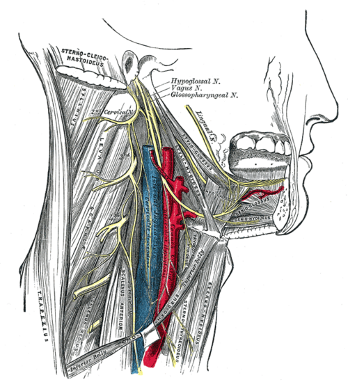
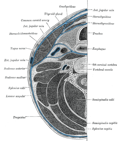
Ascends in the carotid sheath with the internal jugular vein (lateral) and vagus nerve (posterior, between them), under cover of sternocleidomastoid. It bifurcates at the upper border of the thyroid cartilage (about C3–C4) in the carotid triangle (see triangles of the neck). The internal carotid has no branches in the neck; it enters the skull through the carotid canal, runs through the cavernous sinus, and ends by dividing into the anterior and middle cerebral arteries.
- Branches
- Common carotid: no branches in the neck apart from its terminal division
- External carotid artery — face, scalp, neck, dura
- Internal carotid — no neck branches; intracranially the ophthalmic artery (see orbit), posterior communicating, anterior choroidal, then anterior and middle cerebral arteries
- Supplies
Internal carotid — the brain (anterior circulation), eye and orbit, forehead.
- Anastomoses
The circle of Willis links both internal carotids with the vertebrobasilar system (see vertebral artery). Ophthalmic branches anastomose with facial branches of the external carotid — a collateral path if the internal carotid is occluded.
- Relations
The carotid sinus (baroreceptor, CN IX) is a dilation at the origin of the internal carotid; the carotid body (chemoreceptor) lies at the bifurcation. The hypoglossal nerve crosses both carotids above the bifurcation.
- Notes
The carotid pulse is palpated at the anterior border of sternocleidomastoid at the level of the thyroid cartilage.
Branches
Structures supplied
Found in
Mentioned in
- Glossopharyngeal nerve (CN IX)
- Hypoglossal nerve (CN XII)
- Internal jugular vein
- Sternocleidomastoid
- Thyroid gland
Generated automatically from links in other entries.


Clinical correlations
Carotid artery stenosis PANCE
- Mechanism / lesion
Atherosclerotic plaque at the bifurcation/proximal ICA; emboli or low flow cause ischemia in the ICA territory.
- Classic presentation
TIA or stroke — contralateral weakness or numbness, aphasia (dominant hemisphere), or amaurosis fugax (transient monocular blindness, "curtain coming down" — ophthalmic artery emboli); a carotid bruit may be heard. Symptomatic stenosis of 70–99% → carotid endarterectomy (or stenting) within about 2 weeks, plus antiplatelet therapy and statin; 50–69% symptomatic → consider surgery. Asymptomatic stenosis — medical therapy, with selective intervention.
Carotid artery dissection PANCE
- Mechanism / lesion
Intimal tear, spontaneous or after neck trauma or manipulation; a common cause of stroke in young adults.
- Classic presentation
Unilateral neck, face or head pain, ipsilateral Horner syndrome (partial — ptosis and miosis), and TIA/stroke; sometimes pulsatile tinnitus or lower cranial nerve palsies. CTA/MRA diagnoses it.
Carotid sinus hypersensitivity
- Mechanism / lesion
Exaggerated baroreceptor reflex in older adults.
- Classic presentation
Syncope with head turning, tight collars or shaving. Carotid sinus massage (a vagal maneuver) can slow SVT; avoid it if there is a carotid bruit or recent stroke.
High-yield PANCE pearls
- Amaurosis fugax (curtain over one eye) → ICA emboli → carotid duplex.
- Symptomatic 70–99% stenosis → carotid endarterectomy (within 2 weeks).
- Young adult with neck pain + Horner syndrome + stroke → carotid dissection.
- Common carotid bifurcates at the upper border of the thyroid cartilage (C4).
- Carotid sinus = baroreceptor; carotid body = chemoreceptor (both CN IX).
Imaging anatomy
- CT
CT angiography of the head and neck — stenosis, dissection and planning for intervention.
- MRI
MRA (with or without contrast) as an alternative; MRI with fat-saturated sequences shows intramural hematoma in dissection.
- Ultrasound
Carotid duplex ultrasound — first-line to detect and grade stenosis.
- Key views & landmarks
- Carotid duplex (peak systolic velocity grades stenosis)
- CTA head and neck
Physical exam
- Surface landmarks
Anterior border of sternocleidomastoid, level of the thyroid cartilage.
- Palpation
Palpate one carotid at a time (never both together) at the thyroid cartilage level, low in the neck to avoid the carotid sinus. Assess rate, amplitude and upstroke (slow-rising in aortic stenosis).
- Strength / function testing
Auscultate for bruits with the bell while the patient holds a breath. Absence of a bruit does not exclude severe stenosis.
Self-quiz
A 68-year-old man had 10 minutes of painless vision loss in his right eye "like a curtain coming down," which has resolved. What is the most appropriate next test?
Correct answer: B
Amaurosis fugax is a TIA of the retina, usually from emboli from the ipsilateral internal carotid (ophthalmic artery). Urgent carotid imaging (duplex) is needed; consider giant cell arteritis in older patients with headache or jaw claudication.
Show answer
B
Amaurosis fugax is a TIA of the retina, usually from emboli from the ipsilateral internal carotid (ophthalmic artery). Urgent carotid imaging (duplex) is needed; consider giant cell arteritis in older patients with headache or jaw claudication.
A 38-year-old woman has sudden left-sided neck pain after a chiropractic adjustment, followed by right arm weakness. Her left pupil is small and her left eyelid droops. What is the most likely diagnosis?
Correct answer: A
Neck pain + ipsilateral Horner syndrome (sympathetic fibers run on the ICA) + contralateral weakness after neck manipulation indicates left internal carotid dissection.
Show answer
A
Neck pain + ipsilateral Horner syndrome (sympathetic fibers run on the ICA) + contralateral weakness after neck manipulation indicates left internal carotid dissection.
Where does the common carotid artery normally bifurcate?
Correct answer: B
The bifurcation is usually at the upper border of the thyroid cartilage (C3–C4), in the carotid triangle, although it varies.
Show answer
B
The bifurcation is usually at the upper border of the thyroid cartilage (C3–C4), in the carotid triangle, although it varies.
Which cranial nerve carries afferent signals from the carotid sinus baroreceptor?
Correct answer: C
The carotid sinus (baroreceptor, at the origin of the ICA) and the carotid body (chemoreceptor, at the bifurcation) are both supplied by the glossopharyngeal nerve (CN IX).
Show answer
C
The carotid sinus (baroreceptor, at the origin of the ICA) and the carotid body (chemoreceptor, at the bifurcation) are both supplied by the glossopharyngeal nerve (CN IX).
A 70-year-old had a right-hand weakness TIA 5 days ago. Carotid duplex shows 80% stenosis of the left internal carotid artery. What is the most appropriate management?
Correct answer: E
Symptomatic 70–99% ICA stenosis is treated with carotid endarterectomy (or stenting), ideally within about 2 weeks, along with antiplatelet therapy and a statin. A left ICA lesion causes right-sided symptoms.
Show answer
E
Symptomatic 70–99% ICA stenosis is treated with carotid endarterectomy (or stenting), ideally within about 2 weeks, along with antiplatelet therapy and a statin. A left ICA lesion causes right-sided symptoms.