Anal canal
Terminal 3–4 cm of the gut, surrounded by the internal and external anal sphincters. The pectinate line divides it into two halves with different blood supply, lymph drainage, nerves and diseases.
Core anatomy
- Location
In the anal triangle of the perineum, from the anorectal junction (where the rectum passes through the puborectalis sling) to the anus. Surrounded laterally by the ischioanal fossae.
- Parts
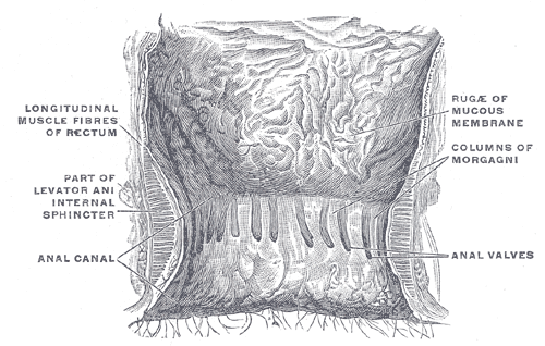
- Anal columns (of Morgagni): longitudinal mucosal ridges containing branches of the superior rectal vessels, joined at their lower ends by the anal valves; behind the valves lie the anal sinuses, into which the anal glands open
- Pectinate (dentate) line: the line of the anal valves; the junction of hindgut endoderm (above) and ectodermal proctodeum (below)
- Anal pecten and anocutaneous line (white line/intersphincteric groove) below the pectinate line; then perianal skin
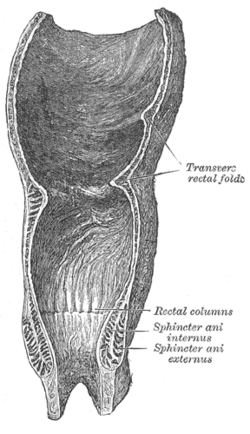
- Internal anal sphincter: involuntary thickening of the circular smooth muscle; provides most resting tone; sympathetic stimulation maintains contraction
- External anal sphincter: voluntary skeletal muscle (subcutaneous, superficial and deep parts), blending with puborectalis above; supplied by the inferior rectal nerve (S4) from the pudendal nerve
- Relations
Anterior: the perineal body and the bulb of the penis/membranous urethra (male) or the lower vagina (female). Posterior: the anococcygeal body and coccyx. Lateral: the ischioanal fossae.
- Arterial supply
Above the pectinate line: superior rectal artery (from the inferior mesenteric artery). Below: inferior rectal arteries (from the internal pudendal artery). Middle rectal arteries anastomose with both.
- Venous drainage
Above: superior rectal vein → IMV → portal system (internal hemorrhoidal plexus). Below: inferior rectal veins → internal pudendal → internal iliac (systemic; external plexus).
- Lymphatic drainage
Above → internal iliac (and inferior mesenteric) nodes. Below → superficial inguinal nodes (see pelvic and inguinal lymph drainage).
- Innervation
Above: visceral (autonomic) — sensitive only to stretch, so lesions are painless. Below: somatic (inferior rectal nerves from the pudendal nerve) — sensitive to pain, touch and temperature, so lesions are painful.
- Notes
Epithelium: columnar above the pectinate line; stratified squamous (non-keratinized in the pecten, keratinized in the perianal skin) below. So adenocarcinoma arises above and squamous cell carcinoma below.
Found in
Mentioned in
Generated automatically from links in other entries.
Swipe sideways to see the whole diagram →


Clinical correlations
Hemorrhoids PANCE
- Mechanism / lesion
Engorgement and descent of the vascular cushions. Internal hemorrhoids arise above the pectinate line (graded I–IV by prolapse); external hemorrhoids below it. Risk: constipation and straining, pregnancy, low-fiber diet.
- Classic presentation
Internal: painless bright-red bleeding on the paper or dripping into the bowl, prolapse. External: perianal swelling; thrombosed external hemorrhoids are acutely painful blue lumps (excise if within about 72 hours).
- Notes
First-line treatment is fiber, fluids and avoiding straining; rubber band ligation for grades I–III internal hemorrhoids.
Anal fissure PANCE
- Mechanism / lesion
Tear in the anoderm below the pectinate line, most often in the posterior midline, usually from passing hard stool, with internal sphincter spasm limiting healing.
- Classic presentation
Severe tearing pain with defecation lasting minutes to hours, and bright-red blood. Chronic fissures have a sentinel skin tag. Fissures that are lateral or multiple suggest Crohn disease, HIV, TB, syphilis or cancer.
- Notes
Fiber and sitz baths, topical nitroglycerin or nifedipine (or diltiazem), botulinum toxin; lateral internal sphincterotomy if refractory.
Anorectal abscess and fistula-in-ano PANCE
- Mechanism / lesion
Infection of an anal gland in an anal sinus (cryptoglandular), spreading into the perianal, ischioanal, intersphincteric or supralevator spaces.
- Classic presentation
Throbbing perianal pain, swelling, fever. Treatment is incision and drainage; a persistent tract after drainage is a fistula. See ischioanal fossa.
Anal cancer
- Mechanism / lesion
Squamous cell carcinoma (below the pectinate line) associated with HPV (16/18), HIV, receptive anal intercourse and immunosuppression.
- Classic presentation
Bleeding, mass, pain; inguinal lymphadenopathy. Treated primarily with chemoradiation (Nigro protocol).
High-yield PANCE pearls
- Pectinate line: above = painless, portal, internal iliac nodes, adenocarcinoma; below = painful, systemic, superficial inguinal nodes, squamous cell carcinoma.
- Painless bright-red bleeding = internal hemorrhoids; exquisitely painful lump = thrombosed external hemorrhoid.
- Tearing pain with defecation + posterior midline tear = anal fissure; off-midline → think Crohn.
- Internal sphincter = involuntary smooth muscle; external = voluntary skeletal (pudendal).
- Anal cancer = SCC, HPV.
Imaging anatomy
- CT
CT pelvis for deep (ischioanal or supralevator) abscesses.
- MRI
Pelvic MRI (fistula protocol) maps complex or recurrent fistulas and abscesses, especially in Crohn disease.
- Ultrasound
Endoanal ultrasound shows sphincter defects (e.g. after obstetric injury) and fistula tracts.
- Key views & landmarks
- Endoanal ultrasound for sphincter integrity
- MRI pelvis fistula protocol
Physical exam
- Surface landmarks
The anal verge; the pectinate line lies about 2 cm above it (seen on anoscopy); the intersphincteric groove is felt as a shallow furrow.
- Palpation
Inspect with the buttocks spread (fissure, tags, thrombosed hemorrhoid, fistula openings). DRE: resting tone (internal sphincter), voluntary squeeze (external sphincter and puborectalis), masses and tenderness. In a suspected fissure, DRE may be deferred because of pain.
- Strength / function testing
Anal wink (anocutaneous reflex, S2–S4): stroking the perianal skin causes contraction of the external sphincter. Anoscopy shows internal hemorrhoids and the pectinate line.
Self-quiz
A 28-year-old woman has severe, tearing anal pain with every bowel movement for 3 weeks, with bright-red blood on the tissue. Inspection shows a longitudinal tear in the posterior midline of the anal verge. What is the most appropriate initial management?
Correct answer: B
This is an acute anal fissure. Initial treatment is conservative: fiber, sitz baths, and topical calcium-channel blockers or nitrates to relax the internal sphincter. Sphincterotomy is for refractory chronic fissures.
Show answer
B
This is an acute anal fissure. Initial treatment is conservative: fiber, sitz baths, and topical calcium-channel blockers or nitrates to relax the internal sphincter. Sphincterotomy is for refractory chronic fissures.
A squamous cell carcinoma arising below the pectinate line would first spread to which lymph nodes?
Correct answer: B
Below the pectinate line, lymph drains to the superficial inguinal nodes, like the perineal skin. Above it, lymph follows the arteries to the internal iliac and inferior mesenteric nodes.
Show answer
B
Below the pectinate line, lymph drains to the superficial inguinal nodes, like the perineal skin. Above it, lymph follows the arteries to the internal iliac and inferior mesenteric nodes.
Which statement about internal hemorrhoids is correct?
Correct answer: C
Internal hemorrhoids arise above the pectinate line, where the mucosa has visceral innervation, so they usually cause painless bleeding. They drain via the superior rectal veins into the portal system.
Show answer
C
Internal hemorrhoids arise above the pectinate line, where the mucosa has visceral innervation, so they usually cause painless bleeding. They drain via the superior rectal veins into the portal system.
An anorectal abscess most often begins as infection of which structure?
Correct answer: C
Most anorectal abscesses are cryptoglandular: an anal gland opening into an anal sinus (behind the anal valves) becomes infected, and the infection spreads into the perianal, ischioanal, intersphincteric or supralevator spaces. Treatment is incision and drainage; a persistent tract afterward is a fistula-in-ano.
Show answer
C
Most anorectal abscesses are cryptoglandular: an anal gland opening into an anal sinus (behind the anal valves) becomes infected, and the infection spreads into the perianal, ischioanal, intersphincteric or supralevator spaces. Treatment is incision and drainage; a persistent tract afterward is a fistula-in-ano.
Which statement about the internal anal sphincter is correct?
Correct answer: E
The internal sphincter is a thickening of the circular smooth muscle, is involuntary, provides most resting tone, and is kept contracted by sympathetic stimulation (it relaxes with parasympathetic activity during defecation). The other statements describe the external sphincter (voluntary, three parts, blends with puborectalis, inferior rectal nerve).
Show answer
E
The internal sphincter is a thickening of the circular smooth muscle, is involuntary, provides most resting tone, and is kept contracted by sympathetic stimulation (it relaxes with parasympathetic activity during defecation). The other statements describe the external sphincter (voluntary, three parts, blends with puborectalis, inferior rectal nerve).
Flashcards
See also
- Rectum
- Ischioanal fossa
- Perineum
- Pudendal nerve
- Anal canal above vs below the pectinate line (comparison table)
- Lymph drainage of the pelvis and perineum (comparison table)