Femoral artery
Continuation of the external iliac artery below the inguinal ligament and the main artery of the lower limb. Its groin pulse is a key exam site and the usual access for catheterization; the superficial femoral segment is the classic site of claudication-causing atherosclerosis.
Core anatomy
- Location
Femoral triangle, then the adductor canal of the thigh.
- Origin
Continuation of the external iliac artery as it passes under the inguinal ligament, at the mid-inguinal point (midway between the ASIS and the pubic symphysis).
- Course
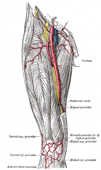
Descends through the femoral triangle (in the femoral sheath, lateral to the femoral vein), enters the adductor canal under sartorius, and passes through the adductor hiatus in adductor magnus to become the popliteal artery.
- Branches
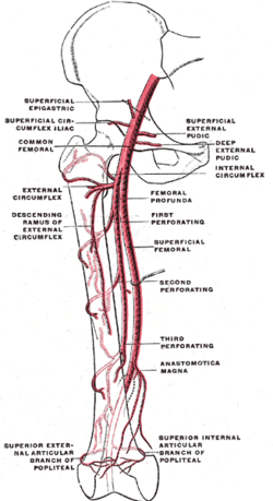
- Superficial branches: superficial epigastric, superficial circumflex iliac, superficial and deep external pudendal
- Deep femoral artery (profunda femoris): about 4 cm below the ligament; gives the medial and lateral circumflex femoral arteries (hip and femoral head) and perforating arteries to the posterior thigh
- Descending genicular artery (to the knee anastomosis)
- Supplies
The lower abdominal wall skin, external genitalia, and the entire lower limb (with the deep femoral supplying most of the thigh).
- Anastomoses
Cruciate and trochanteric anastomoses around the hip (with the gluteal arteries); the genicular anastomosis at the knee — collaterals that open up in chronic occlusion.
- Termination
Becomes the popliteal artery at the adductor hiatus.
- Notes
Clinicians call the segment after the profunda origin the "superficial femoral artery" (SFA) — it is an artery, not a superficial vessel; the "superficial femoral vein" is likewise a deep vein.
Branches
Structures supplied
Found in
Mentioned in
Generated automatically from links in other entries.
Swipe sideways to see the whole diagram →


Clinical correlations
Peripheral artery disease and claudication PANCE
- Mechanism / lesion
Atherosclerosis, most commonly of the superficial femoral artery in the adductor canal; risk factors — smoking, diabetes, hypertension, hyperlipidemia.
- Classic presentation
Calf claudication (reproducible pain with walking, relieved by rest), diminished distal pulses, cool hairless shiny skin. Ankle–brachial index ≤ 0.90 confirms PAD. Treat with smoking cessation, supervised exercise, antiplatelet therapy and a statin; revascularization for limiting symptoms or critical ischemia.
Acute limb ischemia PANCE
- Mechanism / lesion
Embolism (atrial fibrillation) or thrombosis, often lodging at the femoral bifurcation.
- Classic presentation
The 6 Ps: pain, pallor, pulselessness, paresthesia, paralysis, poikilothermia (cold). Emergency — heparin and urgent revascularization; paralysis and sensory loss signal threatened limb.
Femoral access complications PANCE
- Mechanism / lesion
Arterial puncture for angiography, PCI or lines.
- Classic presentation
Groin hematoma, pseudoaneurysm (pulsatile mass with a bruit — ultrasound-guided thrombin injection), AV fistula, or retroperitoneal hemorrhage (high puncture above the inguinal ligament — back/flank pain, hypotension).
High-yield PANCE pearls
- Calf claudication → SFA (adductor canal) disease; buttock claudication → aortoiliac.
- ABI ≤ 0.90 = PAD; > 1.3–1.4 = noncompressible (calcified) vessels.
- 6 Ps = acute limb ischemia → emergency.
- Femoral pulse at the mid-inguinal point; vein is medial to the artery (NAVEL).
- Pulsatile groin mass with bruit after catheterization → pseudoaneurysm.
Imaging anatomy
- CT
CT angiography maps occlusive disease and aneurysms before intervention.
- MRI
MR angiography (alternative without iodinated contrast).
- Ultrasound
ABI and arterial duplex ultrasound first; duplex also diagnoses pseudoaneurysm and AV fistula.
- Key views & landmarks
- Ankle–brachial index
- Arterial duplex of the lower extremity
Physical exam
- Surface landmarks
The femoral pulse is felt just below the mid-inguinal point, halfway between the ASIS and the pubic symphysis.
- Palpation
Compare femoral, popliteal, dorsalis pedis and posterior tibial pulses on both sides; auscultate for femoral bruits.
- Strength / function testing
Inspect for trophic skin changes, capillary refill, Buerger test (elevation pallor, dependent rubor) in chronic ischemia.
Self-quiz
A 64-year-old smoker has cramping pain in his right calf after walking two blocks that resolves with 5 minutes of rest. The right popliteal and pedal pulses are diminished; the femoral pulse is normal. ABI is 0.68. Where is the most likely site of stenosis?
Correct answer: C
Calf claudication with a normal femoral pulse and reduced popliteal and pedal pulses points to the superficial femoral artery, typically in the adductor canal — the most common site of lower-limb PAD.
Show answer
C
Calf claudication with a normal femoral pulse and reduced popliteal and pedal pulses points to the superficial femoral artery, typically in the adductor canal — the most common site of lower-limb PAD.
The femoral head receives most of its blood supply from branches of which vessel?
Correct answer: B
The deep femoral (profunda) artery gives the medial circumflex femoral artery, whose retinacular branches supply most of the femoral head.
Show answer
B
The deep femoral (profunda) artery gives the medial circumflex femoral artery, whose retinacular branches supply most of the femoral head.
A patient with long-standing diabetes has an ankle–brachial index of 1.5. How should this be interpreted?
Correct answer: D
An ABI above about 1.3–1.4 means the tibial arteries are calcified and noncompressible, common in diabetes and CKD, so the ABI can't be interpreted. An ABI ≤ 0.90 confirms PAD.
Show answer
D
An ABI above about 1.3–1.4 means the tibial arteries are calcified and noncompressible, common in diabetes and CKD, so the ABI can't be interpreted. An ABI ≤ 0.90 confirms PAD.
The day after a cardiac catheterization through the right femoral artery, a patient has a pulsatile groin mass with a bruit. What is the most likely diagnosis?
Correct answer: A
A pulsatile mass with a bruit after femoral access is a pseudoaneurysm, usually treated with ultrasound-guided thrombin injection. A simple hematoma isn't expansile or pulsatile; an AV fistula gives a continuous bruit.
Show answer
A
A pulsatile mass with a bruit after femoral access is a pseudoaneurysm, usually treated with ultrasound-guided thrombin injection. A simple hematoma isn't expansile or pulsatile; an AV fistula gives a continuous bruit.
Hours after femoral artery access, a patient develops back and flank pain and hypotension without an obvious groin hematoma. What was the most likely problem with the puncture?
Correct answer: C
A high puncture above the inguinal ligament enters the external iliac artery, where bleeding tracks into the retroperitoneum and can't be compressed, causing back or flank pain and hypotension. Puncture below the ligament allows compression against the femoral head.
Show answer
C
A high puncture above the inguinal ligament enters the external iliac artery, where bleeding tracks into the retroperitoneum and can't be compressed, causing back or flank pain and hypotension. Puncture below the ligament allows compression against the femoral head.