Pulmonary valve
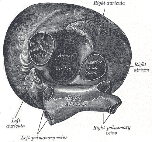
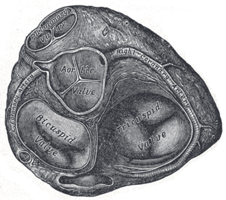
Three-cusped semilunar valve between the right ventricle and the pulmonary trunk. It is the most anterior valve, and the physiologic splitting of S2 is heard best over it.
Core anatomy
- Location
At the top of the conus arteriosus (infundibulum) of the right ventricle, the most anterior and superior of the valves, behind the left 3rd costal cartilage near the sternum. verify
- Parts
- Three semilunar cusps: anterior, right and left
- Pulmonary sinuses above each cusp (no coronary arteries arise from them)
- Relations
Opens into the pulmonary trunk, which lies anterior and to the left of the ascending aorta.
- Notes
Auscultation area: the 2nd left intercostal space at the left sternal border. Physiologic splitting of S2: on inspiration, increased venous return to the right heart delays pulmonic valve closure (P2), so A2 and P2 separate. This is heard best at the pulmonic area.


Clinical correlations
Pulmonic stenosis PANCE
- Mechanism / lesion
Usually congenital. Associated with Noonan syndrome, tetralogy of Fallot, congenital rubella, and carcinoid heart disease.
- Classic presentation
Crescendo–decrescendo systolic ejection murmur at the left upper sternal border that increases with inspiration, sometimes preceded by an ejection click. Wide splitting of S2 (delayed P2). RV heave in severe stenosis.
Abnormal splitting of S2 PANCE
- Mechanism / lesion
Wide split (splitting persists but widens with inspiration): delayed RV emptying (RBBB, pulmonic stenosis). Fixed split (no change with respiration): atrial septal defect. Paradoxical split (splits on expiration): delayed LV emptying (LBBB, severe aortic stenosis).
- Classic presentation
Heard at the pulmonic area; timing with the respiratory cycle distinguishes them.
Pulmonic regurgitation
- Mechanism / lesion
Pulmonary hypertension (dilated annulus) or after repair of tetralogy of Fallot.
- Classic presentation
Early diastolic decrescendo murmur at the left upper sternal border (the Graham Steell murmur in pulmonary hypertension).
High-yield PANCE pearls
- Pulmonic area: left 2nd intercostal space; best place to assess S2 splitting.
- Fixed split S2 = ASD; paradoxical split = LBBB or severe AS; wide split = RBBB or pulmonic stenosis.
- Pulmonic stenosis ↔ Noonan syndrome, tetralogy of Fallot, carcinoid.
Imaging anatomy
- X-ray
Post-stenotic dilation of the pulmonary trunk (prominent pulmonary artery segment on the left heart border) in valvular pulmonic stenosis.
- MRI
Cardiac MRI quantifies pulmonary regurgitation and RV volumes, especially after tetralogy repair.
- Ultrasound
Parasternal short axis at the great-artery level shows the valve and measures the gradient across it (continuous-wave Doppler).
- Key views & landmarks
- Parasternal short axis (aortic valve level) with Doppler across the RV outflow tract
Physical exam
- Surface landmarks
Pulmonic area: left 2nd intercostal space at the sternal border (just below the sternal angle, to the left).
- Palpation
A palpable P2 suggests pulmonary hypertension. A thrill may accompany significant stenosis.
- Strength / function testing
Listen to S2 with the diaphragm during quiet inspiration and expiration to assess splitting.
Self-quiz
A 30-year-old woman has a systolic ejection murmur at the left upper sternal border and a split S2 that does not vary with respiration. What is the most likely diagnosis?
Correct answer: B
A fixed split S2 is characteristic of ASD. The systolic murmur comes from increased flow across the pulmonic valve, not from the defect itself.
Show answer
B
A fixed split S2 is characteristic of ASD. The systolic murmur comes from increased flow across the pulmonic valve, not from the defect itself.
Where is the pulmonic valve best auscultated?
Correct answer: B
The pulmonic area is the left 2nd intercostal space at the sternal border.
Show answer
B
The pulmonic area is the left 2nd intercostal space at the sternal border.
Paradoxical splitting of S2 is most likely associated with which condition?
Correct answer: C
LBBB (and severe AS) delays aortic valve closure, so A2 follows P2 and the split appears on expiration and narrows on inspiration.
Show answer
C
LBBB (and severe AS) delays aortic valve closure, so A2 follows P2 and the split appears on expiration and narrows on inspiration.