Breast
Modified sweat gland on the anterior thoracic wall. Most of its lymph drains to the axillary nodes, which is central to the spread and staging of breast cancer.
Core anatomy
- Location
On the anterior thoracic wall, from about the 2nd to the 6th rib vertically and from the lateral sternal border to the mid-axillary line horizontally. The axillary tail (of Spence) extends toward the axilla.
- Parts
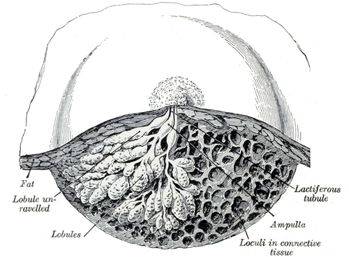
- Glandular tissue arranged in 15–20 lobes, each drained by a lactiferous duct opening on the nipple
- Suspensory ligaments (of Cooper): fibrous bands from the dermis through the gland to the deep fascia
- Retromammary space: loose connective tissue between the breast and the pectoral fascia, allowing the breast to move
- Nipple and areola (areolar glands of Montgomery)
- Relations
Lies on the fascia over pectoralis major, serratus anterior and the external oblique aponeurosis.
- Arterial supply
Medial mammary branches of perforating branches of the internal thoracic artery (the largest supply); lateral thoracic and thoracoacromial arteries (from the axillary artery); posterior intercostal arteries.
- Venous drainage
Mainly to the axillary vein, with tributaries to the internal thoracic and intercostal veins.
- Lymphatic drainage
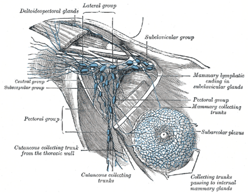
About 75% drains to the axillary nodes, most of it first to the pectoral (anterior) group (see axilla). The medial breast drains mainly to the parasternal nodes along the internal thoracic vessels. Some lymph passes to the opposite breast or to abdominal (inferior phrenic) nodes.
- Innervation
Anterior and lateral cutaneous branches of the 4th–6th intercostal nerves. The nipple is at about the T4 dermatome.
Mentioned in
Generated automatically from links in other entries.


Clinical correlations
Breast cancer PANCE
- Mechanism / lesion
Most are ductal carcinomas. The upper outer quadrant is the most common site (it contains the most glandular tissue). Spreads by lymphatics (axillary nodes first) and blood (bone, lung, liver, brain).
- Classic presentation
A hard, fixed, painless mass. Skin dimpling/retraction (tethering of Cooper ligaments), nipple retraction, peau d'orange (lymphatic obstruction causing edema around hair follicles), bloody nipple discharge, axillary lymphadenopathy.
Paget disease of the nipple PANCE
- Mechanism / lesion
Intraepithelial spread of carcinoma cells (Paget cells) to the nipple epidermis, usually with underlying ductal carcinoma.
- Classic presentation
Eczematous, scaly, crusting or ulcerated nipple that fails to respond to topical treatment. Requires biopsy.
Benign breast conditions PANCE
- Mechanism / lesion
Fibroadenoma (young women): benign fibroepithelial tumor. Fibrocystic changes: hormonally influenced. Mastitis/abscess: usually S. aureus in lactating women. Gynecomastia: glandular proliferation in males (puberty, drugs such as spironolactone, liver disease).
- Classic presentation
Fibroadenoma: firm, rubbery, mobile, well-circumscribed mass. Fibrocystic: cyclic bilateral tenderness and nodularity. Mastitis: painful, red, warm wedge of breast with fever.
Supernumerary nipples (polythelia)
- Mechanism / lesion
Persistence of tissue along the embryonic mammary ridge (milk line) from the axilla to the groin.
- Classic presentation
Small pigmented nodules along the milk line, often mistaken for moles.
High-yield PANCE pearls
- Most common location of breast cancer: upper outer quadrant.
- Peau d'orange = lymphatic obstruction; skin dimpling = Cooper ligament tethering.
- Fibroadenoma: young woman, mobile rubbery mass; ultrasound first in women under ~30.
- Paget disease of the nipple → biopsy; usually underlying carcinoma.
- ~75% of lymph → axillary nodes (pectoral group first); medial breast → parasternal nodes.
Imaging anatomy
- X-ray
Mammography is the main screening test, with two standard views per breast: craniocaudal (CC) and mediolateral oblique (MLO). The MLO includes the axillary tail and low axilla. Suspicious findings: spiculated mass, clustered pleomorphic microcalcifications, architectural distortion. Findings are reported with BI-RADS categories. Screening recommendations (starting age, interval) differ between organizations; USPSTF (2024): mammography every 2 years from age 40 to 74.
- MRI
Used for high-risk screening (e.g. BRCA carriers), extent of disease, implant integrity, and problem-solving.
- Ultrasound
First-line for a palpable mass in women under about 30, pregnant or lactating women, and to tell cystic from solid lesions at any age. Simple cysts are anechoic with posterior acoustic enhancement. Guides core biopsy. Also evaluates axillary nodes.
- Key views & landmarks
- Mammography: craniocaudal (CC) and mediolateral oblique (MLO)
- Targeted ultrasound of a palpable lump
Physical exam
- Surface landmarks
The nipple in males and young women is at about the 4th intercostal space (T4 dermatome); position varies considerably in women.
- Palpation
Inspection with the patient seated: arms at the sides, arms overhead, then hands pressed on the hips (look for asymmetry, dimpling, retraction, skin changes). Palpation with the patient supine and the ipsilateral arm raised: use the pads of the middle three fingers in small circles at three pressures, covering the whole breast systematically (vertical strip pattern) from the clavicle to the inframammary fold and from the sternum to the mid-axillary line, including the axillary tail, nipple and areola. Then examine the axillary, supraclavicular and infraclavicular nodes.
- Strength / function testing
Describe any mass by location (clock face and distance from the nipple), size, shape, consistency, mobility and tenderness.
Self-quiz
A 56-year-old woman has a firm, fixed mass in the upper outer quadrant of the left breast. The overlying skin has a pitted, orange-peel appearance. What causes the skin change?
Correct answer: B
Peau d'orange comes from lymphatic obstruction: dermal edema pulls on hair follicles and pores, producing the pitted appearance. Dimpling and retraction come from tethering of the suspensory (Cooper) ligaments.
Show answer
B
Peau d'orange comes from lymphatic obstruction: dermal edema pulls on hair follicles and pores, producing the pitted appearance. Dimpling and retraction come from tethering of the suspensory (Cooper) ligaments.
Which group of lymph nodes receives most of the lymphatic drainage of the breast?
Correct answer: B
About 75% of breast lymph drains to the axillary nodes, mostly first to the pectoral (anterior) group. The parasternal nodes receive drainage mainly from the medial breast.
Show answer
B
About 75% of breast lymph drains to the axillary nodes, mostly first to the pectoral (anterior) group. The parasternal nodes receive drainage mainly from the medial breast.
A 23-year-old woman has a 2-cm, firm, rubbery, highly mobile, nontender breast mass. What is the most appropriate initial imaging?
Correct answer: B
In women under about 30, ultrasound is the initial test for a palpable mass (dense tissue limits mammography, and radiation dose is a consideration). This presentation suggests a fibroadenoma.
Show answer
B
In women under about 30, ultrasound is the initial test for a palpable mass (dense tissue limits mammography, and radiation dose is a consideration). This presentation suggests a fibroadenoma.
Which artery provides the largest blood supply to the medial breast?
Correct answer: C
Perforating branches of the internal thoracic artery (medial mammary branches) supply the medial breast. The lateral thoracic artery supplies the lateral part.
Show answer
C
Perforating branches of the internal thoracic artery (medial mammary branches) supply the medial breast. The lateral thoracic artery supplies the lateral part.
A breast cancer causes dimpling of the overlying skin. What causes the dimpling?
Correct answer: E
Skin dimpling comes from the tumor tethering Cooper (suspensory) ligaments, which run from the dermis to the deep fascia. Peau d'orange is caused by obstruction of dermal lymphatics; nipple retraction reflects involvement of the ducts behind the nipple.
Show answer
E
Skin dimpling comes from the tumor tethering Cooper (suspensory) ligaments, which run from the dermis to the deep fascia. Peau d'orange is caused by obstruction of dermal lymphatics; nipple retraction reflects involvement of the ducts behind the nipple.