Triangles of the neck
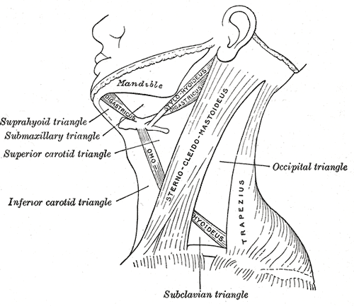
Sternocleidomastoid divides each side of the neck into anterior and posterior triangles, and each is subdivided further. Knowing their contents helps localize neck masses and avoid injuring the accessory nerve.
Core anatomy
- Location
Each side of the neck, divided by sternocleidomastoid.
- Boundaries
- anterior triangle
- Midline of the neck (front), anterior border of SCM (back), inferior border of the mandible (top); apex at the suprasternal notch
- posterior triangle
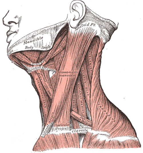
- Posterior border of SCM (front), anterior border of trapezius (back), middle third of the clavicle (base); apex at the occipital bone; roof — investing fascia; floor — prevertebral fascia over splenius, levator scapulae and the scalenes
- submandibular (digastric) triangle
- Mandible and the two bellies of digastric
- carotid triangle
- Superior belly of omohyoid, posterior belly of digastric, anterior border of SCM
- muscular triangle
- Superior belly of omohyoid, anterior border of SCM, midline of the neck
- submental triangle
- Anterior bellies of both digastrics and the hyoid (unpaired)
- occipital and omoclavicular triangles
- Parts of the posterior triangle above and below the inferior belly of omohyoid
- Contents
- Submandibular triangle: Submandibular gland, submandibular nodes, facial artery and vein, hypoglossal nerve
- Carotid triangle: carotid bifurcation, carotid sinus and body (common and internal carotid arteries, external carotid artery), internal jugular vein, vagus and hypoglossal nerves, ansa cervicalis, deep cervical nodes
- Muscular triangle: infrahyoid (strap) muscles, thyroid gland, parathyroid glands, larynx, trachea, esophagus
- Submental triangle: submental nodes, small veins forming the anterior jugular vein
- Posterior triangle: spinal accessory nerve (CN XI) crossing superficially from SCM to trapezius, cutaneous branches of the cervical plexus, external jugular vein, trunks of the brachial plexus, subclavian artery (third part), occipital and supraclavicular nodes
- Communications
The posterior triangle leads down behind the clavicle into the axilla (cervicoaxillary canal) with the brachial plexus and subclavian vessels (see axilla).
- Notes
The hyoid bone (C3) lies at the junction of the submandibular, carotid and muscular triangles.
Mentioned in
- Accessory nerve (CN XI)
- Cervical lymph nodes
- Common and internal carotid arteries
- Sternocleidomastoid
- Submandibular gland
Generated automatically from links in other entries.
Swipe sideways to see the whole diagram →


Clinical correlations
Neck masses by location PANCE
- Mechanism / lesion
Congenital lesions follow embryology; location narrows the differential.
- Classic presentation
Midline: thyroglossal duct cyst (moves with tongue protrusion), dermoid cyst, thyroid isthmus. Anterior triangle (lateral): branchial cleft cyst (2nd arch — along the anterior border of SCM, often after a URI), carotid body tumor (pulsatile, moves side to side but not vertically — Fontaine sign), deep cervical nodes, submandibular gland. Posterior triangle: cystic hygroma (lymphatic malformation, infants, transilluminates), nodes (mono, lymphoma, metastases, TB).
Spinal accessory nerve injury in the posterior triangle PANCE
- Mechanism / lesion
The nerve lies superficially on levator scapulae in the posterior triangle, so it is vulnerable during lymph node biopsy and neck dissection.
- Classic presentation
Trapezius paralysis → shoulder droop, difficulty abducting above 90°, lateral scapular winging; SCM usually spared.
Brachial plexus blocks
- Mechanism / lesion
The trunks of the brachial plexus pass through the lower posterior triangle between the anterior and middle scalenes.
- Classic presentation
Interscalene and supraclavicular blocks are performed here (with ultrasound); nearly all interscalene blocks paralyze the ipsilateral phrenic nerve.
High-yield PANCE pearls
- SCM divides anterior from posterior triangle.
- Posterior triangle node biopsy → CN XI injury → trapezius weakness.
- Lateral neck cyst along the anterior border of SCM → branchial cleft cyst; midline → thyroglossal duct cyst.
- Transilluminating posterior triangle mass in an infant → cystic hygroma (Turner syndrome).
- Carotid triangle → carotid bifurcation (pulse, bruits, carotid body tumor).
Imaging anatomy
- CT
Contrast CT of the neck for adult neck masses and deep infections.
- MRI
Vascular and soft-tissue tumors (e.g. paraganglioma, lymphatic malformation extent).
- Ultrasound
First-line for neck masses in children and for thyroid and nodes; guides FNA.
Physical exam
- Surface landmarks
Turn the head to the opposite side to outline SCM. Hyoid (C3), thyroid cartilage (C4–C5), cricoid (C6); the clavicle forms the base of the posterior triangle.
- Palpation
Palpate each triangle systematically with the lymph node sequence; describe masses by triangle, size, consistency, mobility, pulsatility and relation to swallowing and tongue protrusion.
Self-quiz
A 24-year-old has a smooth, fluctuant, nontender mass along the anterior border of the left sternocleidomastoid that enlarged after a cold. What is the most likely diagnosis?
Correct answer: B
A lateral cystic mass along the anterior border of SCM that swells with URIs is a second branchial cleft cyst. A thyroglossal cyst is midline; a carotid body tumor is firm and pulsatile at the bifurcation.
Show answer
B
A lateral cystic mass along the anterior border of SCM that swells with URIs is a second branchial cleft cyst. A thyroglossal cyst is midline; a carotid body tumor is firm and pulsatile at the bifurcation.
After an excisional biopsy of a node in the posterior triangle, a patient cannot shrug her right shoulder fully and has difficulty lifting her arm above her head. Which nerve was injured?
Correct answer: B
The spinal accessory nerve (CN XI) crosses the posterior triangle superficially and supplies trapezius. Injury causes shoulder droop and weak abduction above 90°.
Show answer
B
The spinal accessory nerve (CN XI) crosses the posterior triangle superficially and supplies trapezius. Injury causes shoulder droop and weak abduction above 90°.
Which structure is found in the carotid triangle?
Correct answer: B
The carotid triangle (omohyoid, posterior digastric, SCM) contains the carotid bifurcation, carotid sinus and body, internal jugular vein, and the vagus and hypoglossal nerves.
Show answer
B
The carotid triangle (omohyoid, posterior digastric, SCM) contains the carotid bifurcation, carotid sinus and body, internal jugular vein, and the vagus and hypoglossal nerves.
A newborn girl with Turner syndrome has a soft, transilluminating mass in the posterior triangle of the neck. What is it most likely to be?
Correct answer: C
A cystic hygroma is a lymphatic malformation, typically a soft, transilluminating posterior triangle mass in an infant, associated with Turner syndrome. Branchial cleft cysts lie along the anterior border of SCM.
Show answer
C
A cystic hygroma is a lymphatic malformation, typically a soft, transilluminating posterior triangle mass in an infant, associated with Turner syndrome. Branchial cleft cysts lie along the anterior border of SCM.
A painless, pulsatile mass at the carotid bifurcation moves side to side but not up and down. What is the most likely diagnosis?
Correct answer: A
A carotid body tumor lies at the bifurcation in the carotid triangle; it is pulsatile and moves horizontally but not vertically (Fontaine sign). Thyroid and thyroglossal lesions move with swallowing.
Show answer
A
A carotid body tumor lies at the bifurcation in the carotid triangle; it is pulsatile and moves horizontally but not vertically (Fontaine sign). Thyroid and thyroglossal lesions move with swallowing.