Extraocular muscles
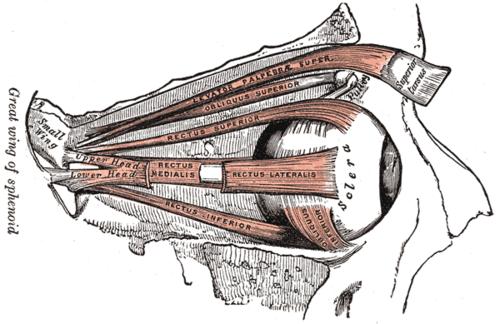
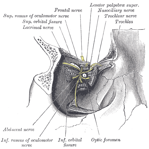
Six muscles that move each eye — four recti and two obliques — supplied by CN III, IV and VI (LR6 SO4, the rest 3). Testing them in the "H" pattern localizes cranial nerve palsies.
Core anatomy
- Location
Within the orbit, around the optic nerve.
- Group
Extraocular muscles of the orbit
- Origin
The four recti arise from the common tendinous ring around the optic canal at the orbital apex. Superior oblique: body of the sphenoid above the ring, passing through the trochlea (a pulley on the superomedial orbital wall). Inferior oblique: floor of the orbit near the nasolacrimal canal (the only one not from the apex).
- Insertion
Recti into the sclera in front of the equator; obliques into the sclera behind the equator (posterolateral), which is why they depress/elevate and rotate.
- Innervation
LR6 SO4, rest 3: lateral rectus — abducens nerve (CN VI); superior oblique — trochlear nerve (CN IV); superior, inferior and medial recti and inferior oblique — oculomotor nerve (CN III, which also supplies levator palpebrae superioris).
- Blood supply
Muscular branches of the ophthalmic artery.
- Action
- Medial rectus: adduction
- Lateral rectus: abduction
- Superior rectus: elevation (mainly when abducted), intorsion, adduction
- Inferior rectus: depression (mainly when abducted), extorsion, adduction
- Superior oblique: depression (mainly when adducted), intorsion, abduction — looking down and in (reading, going downstairs)
- Inferior oblique: elevation (mainly when adducted), extorsion, abduction
- Relations
Clinical (H-pattern) testing isolates each muscle: in abduction, superior rectus elevates and inferior rectus depresses; in adduction, inferior oblique elevates and superior oblique depresses. Levator palpebrae superioris (CN III, with a smooth-muscle part — superior tarsal muscle — supplied by sympathetics) raises the upper lid.
Found in
Mentioned in
Generated automatically from links in other entries.
Swipe sideways to see the whole diagram →


Clinical correlations
Oculomotor (CN III) palsy PANCE
- Mechanism / lesion
Compressive (posterior communicating artery aneurysm, uncal herniation) affects the superficial pupillomotor fibers first → dilated pupil. Ischemic (diabetes, hypertension) spares the pupil.
- Classic presentation
Eye "down and out", ptosis, diplopia; a dilated pupil means compression until proven otherwise (urgent CTA/MRA).
Trochlear (CN IV) palsy PANCE
- Mechanism / lesion
Head trauma (longest intracranial course), congenital.
- Classic presentation
Vertical diplopia worse looking down and in (stairs, reading); the affected eye drifts up; patient tilts the head away from the affected side.
Abducens (CN VI) palsy PANCE
- Mechanism / lesion
Raised intracranial pressure (long course over the petrous apex — a false localizing sign), cavernous sinus lesions, diabetes.
- Classic presentation
Horizontal diplopia worse on looking toward the affected side; the eye can't abduct and rests turned in (esotropia).
Internuclear ophthalmoplegia
- Mechanism / lesion
Lesion of the medial longitudinal fasciculus (multiple sclerosis in the young, stroke in the old).
- Classic presentation
On lateral gaze, the eye on the side of the lesion fails to adduct and the other eye shows nystagmus; convergence is preserved.
High-yield PANCE pearls
- LR6 SO4, rest CN III.
- CN III palsy: down and out + ptosis; dilated pupil = aneurysm (PCOM); pupil-sparing = diabetic/ischemic.
- CN IV palsy: trouble going downstairs, head tilt away from the lesion.
- CN VI palsy: can't abduct; may be a false localizing sign of raised ICP.
- MS in a young adult + failure of adduction with contralateral nystagmus → INO (MLF).
Imaging anatomy
- CT
CT angiography for suspected posterior communicating aneurysm (CN III palsy with dilated pupil); orbital CT for entrapment.
- MRI
MRI brain/orbits for cranial nerve palsies, MS (INO) and thyroid eye disease (enlarged muscle bellies sparing the tendons).
- Key views & landmarks
- CTA/MRA head for pupil-involving CN III palsy
Physical exam
- Surface landmarks
The trochlea is palpable at the superomedial orbital rim.
- Strength / function testing
Hold the head still and trace an "H" with the target: ask about diplopia in each position and look for misalignment and nystagmus. Cover–uncover test for strabismus. Corneal light reflex (Hirschberg) for alignment in children.
Self-quiz
A 58-year-old man has a sudden severe headache. His right eye is deviated down and out with ptosis and a dilated pupil that does not react to light. What is the most likely cause?
Correct answer: B
A pupil-involving CN III palsy suggests compression of the superficial parasympathetic fibers — classically a posterior communicating artery aneurysm, a neurosurgical emergency. Diabetic (ischemic) palsies usually spare the pupil.
Show answer
B
A pupil-involving CN III palsy suggests compression of the superficial parasympathetic fibers — classically a posterior communicating artery aneurysm, a neurosurgical emergency. Diabetic (ischemic) palsies usually spare the pupil.
To test the superior oblique in isolation, the patient should look in which direction?
Correct answer: C
With the eye adducted, the superior oblique becomes the main depressor, so looking down and in (toward the nose) tests it — the position used for reading or walking downstairs.
Show answer
C
With the eye adducted, the superior oblique becomes the main depressor, so looking down and in (toward the nose) tests it — the position used for reading or walking downstairs.
Which nerve supplies the lateral rectus?
Correct answer: C
The abducens nerve (CN VI) supplies the lateral rectus (LR6). CN IV supplies the superior oblique; CN III supplies the rest.
Show answer
C
The abducens nerve (CN VI) supplies the lateral rectus (LR6). CN IV supplies the superior oblique; CN III supplies the rest.
A patient with a right trochlear (CN IV) palsy tilts the head in which direction to reduce diplopia?
Correct answer: B
In CN IV palsy the affected eye drifts up and extorts; patients tilt the head away from the affected side to compensate. Diplopia is worst looking down and in (reading, going downstairs).
Show answer
B
In CN IV palsy the affected eye drifts up and extorts; patients tilt the head away from the affected side to compensate. Diplopia is worst looking down and in (reading, going downstairs).
Which extraocular muscle does NOT arise from the orbital apex?
Correct answer: E
The inferior oblique arises from the orbital floor near the nasolacrimal canal. The four recti arise from the common tendinous ring, and the superior oblique from the sphenoid above it, passing through the trochlea.
Show answer
E
The inferior oblique arises from the orbital floor near the nasolacrimal canal. The four recti arise from the common tendinous ring, and the superior oblique from the sphenoid above it, passing through the trochlea.