Cubital fossa
Triangular depression on the anterior elbow containing the biceps tendon, brachial artery and median nerve. It is the usual site for venipuncture and blood pressure auscultation.
Core anatomy
- Location
Anterior aspect of the elbow.
- Boundaries
- superior
- Imaginary line joining the medial and lateral epicondyles of the humerus
- medial
- Pronator teres
- lateral
- Brachioradialis
- apex
- Where the medial and lateral boundaries meet, distally
- floor
- Brachialis (proximally) and supinator (distally)
- roof
- Skin, subcutaneous tissue (with the superficial veins and cutaneous nerves), deep fascia, and the bicipital aponeurosis
- Contents
- Biceps brachii tendon
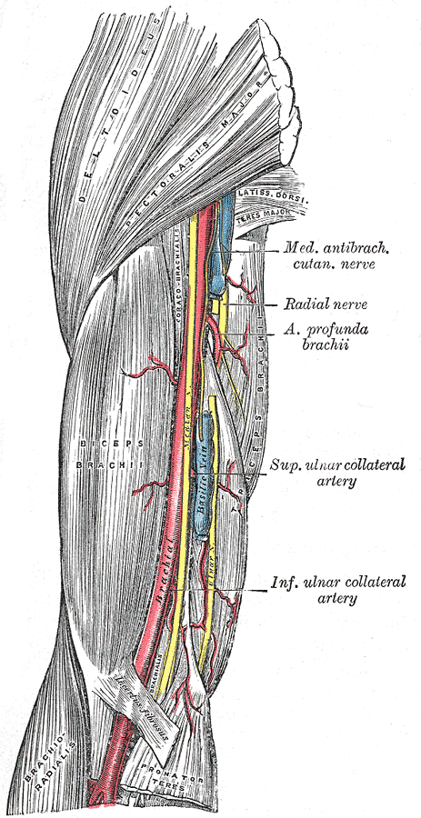
- Brachial artery, dividing at the apex into the radial artery and ulnar artery
- Median nerve (most medial)
- Radial nerve: deep, at the lateral edge between brachialis and brachioradialis, dividing into superficial and deep branches
- Communications
Continuous with the anterior compartment of the arm above and the anterior compartment of the forearm below.
- Notes
Main contents from lateral to medial are remembered as TAN: biceps Tendon, brachial Artery, median Nerve.
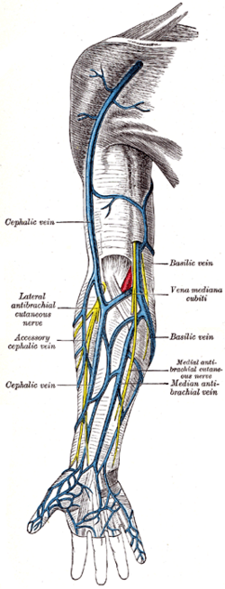
Superficial veins in the roof: the cephalic vein (lateral) and basilic vein (medial) are connected by the median cubital vein, which crosses superficial to the bicipital aponeurosis. The aponeurosis separates the vein from the brachial artery and median nerve. The superficial vein pattern varies considerably between people.
Mentioned in
- Biceps brachii
- Brachial artery
- Elbow joint
- Median nerve
- Pronator teres
- Radial artery
- Radial nerve
- Ulnar artery
Generated automatically from links in other entries.
Swipe sideways to see the whole diagram →


Clinical correlations
Venipuncture and IV access PANCE
- Mechanism / lesion
The median cubital vein is the preferred site because it is superficial, large and anchored.
- Classic presentation
The bicipital aponeurosis provides some protection to the brachial artery and median nerve beneath it. Risks include hematoma, puncture of the brachial artery (bright red, pulsatile return), and injury to cutaneous nerves (lateral or medial cutaneous nerves of the forearm).
Supracondylar fracture PANCE
- Mechanism / lesion
Displacement of the distal humerus fragments into the fossa.
- Classic presentation
Risk to the brachial artery and median nerve/AIN with swelling that may progress to compartment syndrome.
Distal biceps tendon rupture
- Mechanism / lesion
Eccentric loading of the flexed elbow.
- Classic presentation
Loss of the palpable tendon in the fossa (positive hook test), bruising and weak supination.
High-yield PANCE pearls
- Cubital fossa contents, lateral → medial: biceps Tendon, brachial Artery, median Nerve (TAN); radial nerve deep at the lateral edge.
- Median cubital vein → venipuncture; superficial to the bicipital aponeurosis.
- Brachial pulse / BP auscultation: medial to the biceps tendon.
Imaging anatomy
- X-ray
The fossa is a soft-tissue region. Elbow radiographs show fat pads and fractures.
- MRI
Distal biceps tendon (FABS view), brachialis injury, and masses.
- Ultrasound
Ultrasound-guided IV access in difficult patients (identify the vein and avoid the artery and nerve). Assessment of the distal biceps tendon and the brachial artery bifurcation.
- Key views & landmarks
- Transverse ultrasound across the fossa showing biceps tendon → artery → median nerve (lateral to medial)
Physical exam
- Surface landmarks
The biceps tendon is felt as a firm cord when the patient flexes the supinated forearm against resistance. The brachial pulse is just medial to it. The superficial veins become visible with a tourniquet.
- Palpation
Palpate the biceps tendon, the brachial pulse and the medial and lateral borders (pronator teres, brachioradialis).
- Strength / function testing
Hook test for distal biceps integrity. BP auscultation over the brachial artery.
Self-quiz
During venipuncture in the antecubital fossa, the needle is advanced too deeply through the median cubital vein medial to the biceps tendon. Which structure is most at risk?
Correct answer: B
Deep to the bicipital aponeurosis, medial to the biceps tendon, lies the brachial artery, with the median nerve medial to it. The ulnar nerve is behind the medial epicondyle, not in the fossa.
Show answer
B
Deep to the bicipital aponeurosis, medial to the biceps tendon, lies the brachial artery, with the median nerve medial to it. The ulnar nerve is behind the medial epicondyle, not in the fossa.
What is the correct order of the main cubital fossa contents from lateral to medial?
Correct answer: B
Lateral to medial: biceps Tendon, brachial Artery, median Nerve (TAN). The radial nerve lies deep at the lateral margin, under brachioradialis.
Show answer
B
Lateral to medial: biceps Tendon, brachial Artery, median Nerve (TAN). The radial nerve lies deep at the lateral margin, under brachioradialis.
Which structure forms the medial boundary of the cubital fossa?
Correct answer: C
Pronator teres forms the medial border and brachioradialis the lateral border. Brachialis and supinator form the floor.
Show answer
C
Pronator teres forms the medial border and brachioradialis the lateral border. Brachialis and supinator form the floor.
Which muscle forms the lateral boundary of the cubital fossa?
Correct answer: E
The cubital fossa is bounded laterally by brachioradialis, medially by pronator teres, and superiorly by a line between the epicondyles. Brachialis and supinator form its floor.
Show answer
E
The cubital fossa is bounded laterally by brachioradialis, medially by pronator teres, and superiorly by a line between the epicondyles. Brachialis and supinator form its floor.
Which structure separates the median cubital vein from the brachial artery and median nerve?
Correct answer: A
The bicipital aponeurosis passes over the brachial artery and median nerve, and the median cubital vein lies superficial to it. This gives some protection during venipuncture, but a needle passed too deeply medial to the biceps tendon can still reach the artery.
Show answer
A
The bicipital aponeurosis passes over the brachial artery and median nerve, and the median cubital vein lies superficial to it. This gives some protection during venipuncture, but a needle passed too deeply medial to the biceps tendon can still reach the artery.