Cruciate ligaments
Crossed intra-articular ligaments of the knee that prevent the tibia sliding forward (ACL) or backward (PCL) on the femur. ACL tears from non-contact pivoting are among the most common serious sports injuries.
Core anatomy
- Location
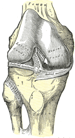
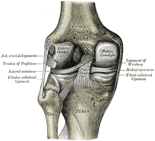
Inside the capsule of the knee joint but outside its synovial cavity, in the intercondylar fossa, crossing like an X.
- Attachments
ACL: from the anterior intercondylar area of the tibia upward, backward and laterally to the medial side of the lateral femoral condyle. PCL: from the posterior intercondylar area of the tibia upward, forward and medially to the lateral side of the medial femoral condyle. (Named for their tibial attachments.)
- Function
ACL: prevents anterior translation of the tibia on the femur (posterior displacement of the femur on the tibia) and limits hyperextension and rotation; it is the weaker of the two. PCL: prevents posterior translation of the tibia; the stronger, main stabilizer when weight-bearing on a flexed knee (walking downhill).
- Relations
Supplied by the middle genicular artery (from the popliteal artery). The ACL is often torn together with the MCL and medial meniscus.
Swipe sideways to see the whole diagram →


Clinical correlations
ACL tear PANCE
- Mechanism / lesion
Usually non-contact: sudden deceleration, cutting or pivoting, or landing from a jump with the knee near extension and in valgus. More common in female athletes. Contact valgus blows can tear the "unhappy triad" (ACL, MCL and medial meniscus; the lateral meniscus is often involved in practice).
- Classic presentation
A "pop", immediate inability to continue, and rapid hemarthrosis (within hours); later instability ("giving way") with pivoting. Positive Lachman (most sensitive), anterior drawer and pivot-shift tests.
- Notes
Confirm with MRI. Young active patients usually have ACL reconstruction; others may do well with rehabilitation alone.
PCL tear PANCE
- Mechanism / lesion
A posteriorly directed force on the proximal tibia with the knee flexed — dashboard injury or a fall onto the flexed knee.
- Classic presentation
Posterior sag of the tibia, positive posterior drawer test; often less disabling than ACL tears and frequently managed non-operatively when isolated.
High-yield PANCE pearls
- Non-contact pivot + pop + rapid swelling → ACL tear → Lachman (most sensitive).
- Dashboard injury to the flexed knee → PCL tear (posterior drawer, sag sign).
- Unhappy triad: ACL + MCL + medial meniscus.
- ACL prevents anterior tibial translation; PCL prevents posterior.
Imaging anatomy
- X-ray
Usually normal; a Segond fracture (avulsion of the lateral tibial plateau) is strongly associated with ACL tears. Rule out fractures (Ottawa knee rules).
- MRI
MRI confirms the tear and shows associated injuries — the "kissing" bone bruises of the lateral femoral condyle and posterolateral tibial plateau, meniscal and MCL tears.
- Key views & landmarks
- MRI knee (sagittal proton-density)
Physical exam
- Surface landmarks
Not palpable; tested by stress maneuvers comparing both knees.
- Strength / function testing
Examine soon after injury (before guarding) or after the swelling settles; always compare with the uninjured side.
Lachman test
- Technique
Knee flexed 20–30°; stabilize the distal femur with one hand and pull the proximal tibia forward with the other.
- Positive test
Increased anterior translation with a soft or absent end point compared with the other side.
- Significance
The most sensitive test for ACL tear.
Anterior drawer test
- Technique
Knee flexed 90° with the foot stabilized (sit on it); pull the proximal tibia forward.
- Positive test
Excess anterior translation.
- Significance
ACL tear; less sensitive than Lachman in acute injuries (hamstring guarding).
Pivot-shift test
- Technique
With the knee extended, apply valgus and internal rotation, then flex the knee.
- Positive test
A clunk as the subluxed lateral tibial plateau reduces at about 20–40° of flexion.
- Significance
The most specific test for ACL insufficiency, but hard to do in an awake patient.
Posterior drawer test and sag sign
- Technique
Knee at 90°: push the proximal tibia backward. Sag sign: with hips and knees flexed to 90°, look from the side.
- Positive test
Excess posterior translation; the tibia sags backward under gravity.
- Significance
PCL tear.
Self-quiz
A 19-year-old female soccer player plants her foot and pivots, feels a pop, and falls. Her knee swells within 2 hours. Which exam test is most sensitive for the most likely injury?
Correct answer: B
A non-contact pivoting injury with a pop and rapid hemarthrosis suggests an ACL tear. The Lachman test at 20–30° of flexion is the most sensitive exam maneuver.
Show answer
B
A non-contact pivoting injury with a pop and rapid hemarthrosis suggests an ACL tear. The Lachman test at 20–30° of flexion is the most sensitive exam maneuver.
A driver hits his flexed knee on the dashboard. The tibia sags backward when both knees are flexed to 90°. Which structure is torn?
Correct answer: B
A dashboard injury drives the tibia backward, tearing the PCL; the posterior sag sign and posterior drawer test are positive.
Show answer
B
A dashboard injury drives the tibia backward, tearing the PCL; the posterior sag sign and posterior drawer test are positive.
The ACL attaches to the femur on which surface?
Correct answer: B
The ACL runs from the anterior tibial intercondylar area to the medial side of the lateral femoral condyle; the PCL runs to the lateral side of the medial condyle.
Show answer
B
The ACL runs from the anterior tibial intercondylar area to the medial side of the lateral femoral condyle; the PCL runs to the lateral side of the medial condyle.
Which ligament is the main stabilizer when weight-bearing on a flexed knee, as when walking downhill?
Correct answer: A
The PCL is the stronger cruciate and prevents posterior translation of the tibia (anterior sliding of the femur on the tibia), which matters most when bearing weight on a flexed knee. The ACL prevents anterior tibial translation.
Show answer
A
The PCL is the stronger cruciate and prevents posterior translation of the tibia (anterior sliding of the femur on the tibia), which matters most when bearing weight on a flexed knee. The ACL prevents anterior tibial translation.
MRI confirms a complete ACL tear in a 20-year-old competitive soccer player who wants to return to pivoting sports. What is the usual management?
Correct answer: E
Young, active patients who pivot usually have ACL reconstruction; less active patients may do well with rehabilitation alone. Immobilization and injection don't restore stability.
Show answer
E
Young, active patients who pivot usually have ACL reconstruction; less active patients may do well with rehabilitation alone. Immobilization and injection don't restore stability.
Flashcards
See also
- Knee joint
- Collateral ligaments of the knee
- Menisci of the knee
- Knee ligament and meniscal injuries (comparison table)
- A pop in the knee on landing (clinical case)