Lesser sac
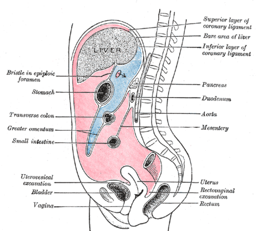
Part of the peritoneal cavity behind the stomach, entered through the omental (epiploic) foramen. The hepatoduodenal ligament in front of that foramen is compressed in the Pringle maneuver.
Core anatomy
- Location
Posterior to the stomach and lesser omentum, anterior to the pancreas, left kidney and suprarenal gland.
- Boundaries
- anterior
- Lesser omentum, posterior surface of the stomach, anterior layers of the greater omentum
- posterior
- Pancreas, left kidney and suprarenal gland (covered by peritoneum), aorta
- superior
- Caudate lobe of the liver and diaphragm
- inferior
- Fused layers of the greater omentum / transverse mesocolon
- left
- Spleen and the gastrosplenic and splenorenal ligaments
- Contents
- A potential space normally containing only a thin film of fluid
- Communications
Connects with the greater sac through the omental (epiploic) foramen (of Winslow). Its boundaries are: anterior, the free edge of the hepatoduodenal ligament (containing the portal triad: Hepatic portal vein posteriorly, hepatic artery proper on the left, common bile duct on the right); posterior, the inferior vena cava; superior, the caudate lobe of the liver; inferior, the first part of the duodenum.
Found in
Mentioned in
Generated automatically from links in other entries.

Clinical correlations
Pringle maneuver PANCE
- Mechanism / lesion
Compressing the hepatoduodenal ligament (index finger in the omental foramen, thumb in front) occludes the hepatic artery and portal vein.
- Classic presentation
Used to control hepatic bleeding. If bleeding continues despite the maneuver, the source is likely the hepatic veins or retrohepatic IVC (which aren't controlled by it).
Collections and perforations in the lesser sac PANCE
- Mechanism / lesion
Pancreatic pseudocysts and fluid from pancreatitis collect here. A perforated posterior gastric ulcer leaks into the lesser sac and can erode the pancreas or splenic artery.
- Classic presentation
Epigastric fullness, early satiety or pain. A retrogastric collection on CT.
High-yield PANCE pearls
- Portal triad in the hepatoduodenal ligament: portal vein (posterior), hepatic artery (left), CBD (right).
- Pringle maneuver controls inflow; persistent bleeding → hepatic veins/IVC.
- Pancreatic pseudocyst classically sits in the lesser sac (behind the stomach).
Imaging anatomy
- CT
Shows retrogastric fluid collections and pseudocysts (well-defined wall, typically 4 or more weeks after acute pancreatitis) and their relation to the stomach.
- Ultrasound
Pseudocysts may be seen through the left lobe of the liver or stomach. Endoscopic ultrasound guides drainage into the stomach (cystgastrostomy).
- Key views & landmarks
- CT abdomen with contrast
Physical exam
- Palpation
Large pseudocysts may present as an epigastric mass.
Self-quiz
Which structure forms the posterior boundary of the omental (epiploic) foramen?
Correct answer: B
The IVC lies behind the omental foramen. The hepatoduodenal ligament is anterior, the caudate lobe superior, and the first part of the duodenum inferior.
Show answer
B
The IVC lies behind the omental foramen. The hepatoduodenal ligament is anterior, the caudate lobe superior, and the first part of the duodenum inferior.
During a trauma laparotomy, a surgeon compresses the hepatoduodenal ligament, but brisk bleeding continues from behind the liver. What is the most likely source?
Correct answer: C
The Pringle maneuver occludes the hepatic artery and portal vein (inflow). Continued bleeding suggests injury to the hepatic veins or retrohepatic IVC, which drain the liver (outflow).
Show answer
C
The Pringle maneuver occludes the hepatic artery and portal vein (inflow). Continued bleeding suggests injury to the hepatic veins or retrohepatic IVC, which drain the liver (outflow).
Within the hepatoduodenal ligament, which structure lies most posteriorly?
Correct answer: C
The portal vein is posterior, with the common bile duct to the right and the hepatic artery proper to the left anteriorly.
Show answer
C
The portal vein is posterior, with the common bile duct to the right and the hepatic artery proper to the left anteriorly.