Internal capsule
Compact band of white matter carrying motor and sensory fibers between the cortex and the brainstem. A small (lacunar) infarct here can cause a pure motor hemiparesis of the face, arm and leg.
Core anatomy
- Location
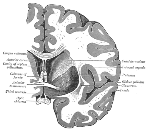
Deep in each hemisphere, V-shaped in horizontal section, between the caudate and thalamus (medially) and the lentiform nucleus (laterally) — see basal ganglia, thalamus.
- Parts
- Anterior limb (between caudate and lentiform): frontopontine and thalamocortical (anterior/mediodorsal) fibers
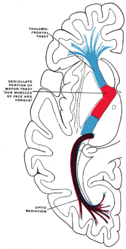
- Genu: corticobulbar fibers (face, head)
- Posterior limb (between thalamus and lentiform): corticospinal fibers (arm anterior, leg posterior) and thalamocortical sensory radiations (from VPL/VPM)
- Retrolenticular and sublenticular parts: optic radiations and auditory radiations
- Continues below as the cerebral peduncle of the midbrain and above as the corona radiata
- Relations
Because motor fibers for the whole half-body are packed tightly together, a small lesion causes a large deficit — contralateral face, arm and leg equally.
- Arterial supply
Lenticulostriate arteries (from the MCA) — most of the capsule; anterior choroidal artery (from the ICA) — lower posterior limb; recurrent artery of Heubner (from the ACA) — anterior limb and genu. See cerebral arteries (ACA, MCA, PCA).
- Notes
Contains the corticospinal tract and the sensory projections from the thalamus.
Mentioned in
Generated automatically from links in other entries.


Clinical correlations
Lacunar infarcts PANCE
- Mechanism / lesion
Occlusion of a single deep penetrating artery (lipohyalinosis) from chronic hypertension and diabetes; infarcts under 1.5 cm.
- Classic presentation
Classic syndromes, without cortical signs (no aphasia, neglect or visual field loss): pure motor hemiparesis (posterior limb or basis pontis — contralateral face, arm and leg equally); pure sensory stroke (thalamus); sensorimotor stroke (thalamus + capsule); ataxic hemiparesis and dysarthria–clumsy hand syndrome (capsule or pons).
Hypertensive intracerebral hemorrhage PANCE
- Mechanism / lesion
Rupture of lenticulostriate vessels in the putamen or thalamus extending into the capsule.
- Classic presentation
Sudden contralateral hemiplegia and sensory loss, headache, vomiting, declining consciousness; very high blood pressure. Noncontrast CT; lower BP, reverse anticoagulation.
High-yield PANCE pearls
- Pure motor hemiparesis of face, arm and leg equally, no cortical signs → lacunar infarct of the posterior limb.
- Lacunes → hypertension and diabetes (small vessel disease).
- Cortical signs (aphasia, neglect, field cut, gaze deviation) → cortical (large vessel) stroke, not lacunar.
Imaging anatomy
- CT
Noncontrast CT excludes hemorrhage; small acute lacunes are often invisible early.
- MRI
DWI MRI shows acute lacunar infarcts; T2/FLAIR shows chronic small vessel disease (white matter hyperintensities, old lacunes).
Physical exam
- Strength / function testing
Compare face, arm and leg strength (equal involvement suggests capsular lesion; face/arm > leg suggests MCA cortex); check for cortical signs — language, neglect, visual fields, cortical sensation; upper motor neuron signs (hyperreflexia, Babinski).
Self-quiz
A 66-year-old with long-standing hypertension develops weakness of the left face, arm and leg to the same degree. Sensation, language, visual fields and attention are normal. Where is the most likely lesion?
Correct answer: B
Pure motor hemiparesis of the face, arm and leg equally, with no cortical signs, is a lacunar infarct of the posterior limb of the internal capsule (lenticulostriate branch of the MCA).
Show answer
B
Pure motor hemiparesis of the face, arm and leg equally, with no cortical signs, is a lacunar infarct of the posterior limb of the internal capsule (lenticulostriate branch of the MCA).
Which fibers pass through the genu of the internal capsule?
Correct answer: B
The genu carries corticobulbar fibers to cranial nerve motor nuclei. Corticospinal fibers and sensory radiations are in the posterior limb; optic radiations are retrolenticular.
Show answer
B
The genu carries corticobulbar fibers to cranial nerve motor nuclei. Corticospinal fibers and sensory radiations are in the posterior limb; optic radiations are retrolenticular.
Which risk factors are most associated with lacunar infarcts of the internal capsule?
Correct answer: B
Lacunes are small-vessel infarcts from lipohyalinosis of deep penetrating arteries, caused mainly by chronic hypertension and diabetes. Atrial fibrillation causes embolic cortical strokes.
Show answer
B
Lacunes are small-vessel infarcts from lipohyalinosis of deep penetrating arteries, caused mainly by chronic hypertension and diabetes. Atrial fibrillation causes embolic cortical strokes.
Which finding argues against a lacunar infarct?
Correct answer: E
Lacunar syndromes lack cortical signs: aphasia, neglect, visual field loss and gaze deviation point to a cortical (large-vessel) stroke.
Show answer
E
Lacunar syndromes lack cortical signs: aphasia, neglect, visual field loss and gaze deviation point to a cortical (large-vessel) stroke.
Which vessels supply most of the internal capsule?
Correct answer: A
Most of the internal capsule is supplied by the lenticulostriate arteries of the MCA, with contributions from the anterior choroidal artery (posterior limb) and the recurrent artery of Heubner (anterior limb and genu).
Show answer
A
Most of the internal capsule is supplied by the lenticulostriate arteries of the MCA, with contributions from the anterior choroidal artery (posterior limb) and the recurrent artery of Heubner (anterior limb and genu).
Flashcards
See also
- Basal ganglia
- Thalamus
- Corticospinal tract
- Cerebral arteries (ACA, MCA, PCA)
- Cerebral cortex and lobes
- Intracranial hemorrhage (comparison table)
- Stroke syndromes by territory (comparison table)
- Sudden aphasia and arm weakness (clinical case)