Cardiac conduction system
Specialized cardiac muscle that generates and conducts impulses (SA node → AV node → bundle of His → bundle branches → Purkinje fibers). Its blood supply explains the arrhythmias that accompany some MIs.
Core anatomy
- Location
Within the walls of the heart.
- Parts
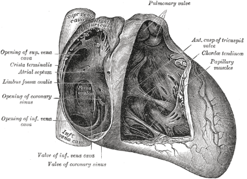
- Sinoatrial (SA) node: the pacemaker, in the wall of the right atrium near the opening of the superior vena cava, at the upper end of the crista terminalis
- Atrioventricular (AV) node: in the interatrial septum near the opening of the coronary sinus (triangle of Koch). Delays conduction so the ventricles can fill
- AV bundle (of His): passes through the fibrous skeleton and along the membranous interventricular septum, the only normal electrical connection between atria and ventricles
- Right and left bundle branches: descend on each side of the muscular septum. The right branch continues in the septomarginal trabecula (moderator band)
- Subendocardial (Purkinje) fibers: spread the impulse through the ventricular myocardium
- Arterial supply
SA nodal artery: usually from the right coronary artery (about 60%), otherwise from the circumflex artery. AV nodal artery: from the dominant coronary artery, usually the RCA (about 80%). Bundle branches: septal perforators of the anterior interventricular artery (anterior two-thirds of the septum) and of the posterior interventricular artery. verify
- Innervation
Sympathetic fibers increase rate and conduction velocity. Parasympathetic (vagus) fibers slow the SA node and AV conduction; the right vagus predominantly affects the SA node and the left vagus the AV node. verify

Clinical correlations
Bradyarrhythmias and AV block in inferior MI PANCE
- Mechanism / lesion
The RCA usually supplies the SA and AV nodes, so inferior MI (RCA occlusion) can cause sinus bradycardia and AV block (often transient, at the AV node).
- Classic presentation
Bradycardia and hypotension with ST elevation in II, III and aVF. First-degree or Mobitz I block is typical at the AV node level.
Bundle branch block PANCE
- Mechanism / lesion
Conduction delay in the right or left bundle branch (ischemia, fibrosis, hypertrophy, or after surgery).
- Classic presentation
Wide QRS (120 ms or more). RBBB: rSR' in V1–V2, wide S in I and V6. LBBB: broad notched R in I, aVL, V5–V6; deep S in V1. A new LBBB with symptoms of ischemia needs evaluation for acute MI.
Pre-excitation (Wolff–Parkinson–White) PANCE
- Mechanism / lesion
An accessory pathway (bundle of Kent) bypasses the AV node and connects atrium to ventricle directly.
- Classic presentation
Short PR interval, delta wave, wide QRS. Predisposes to reentrant tachycardias. In atrial fibrillation with WPW, AV nodal blockers (adenosine, beta blockers, calcium channel blockers, digoxin) can be dangerous; procainamide or cardioversion is used.
Sick sinus syndrome
- Mechanism / lesion
Degenerative dysfunction of the SA node, usually in older adults.
- Classic presentation
Sinus bradycardia, pauses, or alternating bradycardia and tachycardia (tachy-brady syndrome), with fatigue, dizziness or syncope. Often treated with a pacemaker.
High-yield PANCE pearls
- Conduction: SA → AV (delay) → His → bundle branches → Purkinje.
- Inferior MI (RCA) → bradycardia / AV block (the RCA usually supplies the SA and AV nodes).
- WPW: short PR + delta wave; avoid AV nodal blockers in pre-excited AF.
- New LBBB with chest pain → treat as a possible acute MI.
Imaging anatomy
- X-ray
The system is not visible. Pacemaker and ICD leads are seen on CXR (RA lead in the appendage, RV lead at the apex or septum).
- MRI
Cardiac MRI can show infiltrative disease (sarcoidosis, amyloidosis) causing conduction disease.
- Ultrasound
Echo assesses structural causes of arrhythmia (valve disease, cardiomyopathy, LA size).
- Key views & landmarks
- 12-lead ECG: the functional image of the conduction system (PR interval = AV conduction; QRS width = ventricular conduction)
Physical exam
- Strength / function testing
Pulse rate and regularity. Irregularly irregular pulse = atrial fibrillation until proven otherwise. Cannon a waves in the JVP suggest AV dissociation (complete heart block). Variable intensity of S1 also occurs in complete heart block.
Self-quiz
A 62-year-old man has ST elevation in leads II, III and aVF. His heart rate is 42/min, and the ECG shows a PR interval that lengthens progressively until a QRS is dropped. Which artery is most likely occluded?
Correct answer: C
Inferior ST elevation with Mobitz I (Wenckebach) block points to the RCA, which usually supplies the AV node (right-dominant circulation).
Show answer
C
Inferior ST elevation with Mobitz I (Wenckebach) block points to the RCA, which usually supplies the AV node (right-dominant circulation).
Where is the sinoatrial node located?
Correct answer: B
The SA node lies in the right atrial wall at the junction with the SVC, at the upper end of the crista terminalis. The AV node lies near the coronary sinus opening in the interatrial septum.
Show answer
B
The SA node lies in the right atrial wall at the junction with the SVC, at the upper end of the crista terminalis. The AV node lies near the coronary sinus opening in the interatrial septum.
An ECG shows a short PR interval, a slurred upstroke of the QRS, and a QRS duration of 125 ms. What is the underlying abnormality?
Correct answer: B
A short PR with a delta wave and wide QRS is Wolff–Parkinson–White pre-excitation: an accessory pathway (bundle of Kent) bypasses the AV node.
Show answer
B
A short PR with a delta wave and wide QRS is Wolff–Parkinson–White pre-excitation: an accessory pathway (bundle of Kent) bypasses the AV node.
Which structure carries the right bundle branch across the right ventricular cavity to the anterior papillary muscle?
Correct answer: B
The septomarginal trabecula (moderator band) carries part of the right bundle branch from the septum to the base of the anterior papillary muscle, shortening conduction time.
Show answer
B
The septomarginal trabecula (moderator band) carries part of the right bundle branch from the septum to the base of the anterior papillary muscle, shortening conduction time.
A 30-year-old with known Wolff–Parkinson–White syndrome presents with an irregular, wide-complex tachycardia at 210/min (pre-excited atrial fibrillation). He is hemodynamically stable. Which drug is appropriate?
Correct answer: C
In pre-excited AF, AV nodal blockers (adenosine, calcium channel blockers, beta blockers, digoxin) can increase conduction down the accessory pathway and precipitate ventricular fibrillation. Procainamide (or cardioversion if unstable) is used.
Show answer
C
In pre-excited AF, AV nodal blockers (adenosine, calcium channel blockers, beta blockers, digoxin) can increase conduction down the accessory pathway and precipitate ventricular fibrillation. Procainamide (or cardioversion if unstable) is used.
Flashcards
See also
- Heart
- Right coronary artery
- Chest pain with a slow pulse (clinical case)