Menisci of the knee
Two crescent-shaped fibrocartilage pads on the tibial plateau that deepen the articular surfaces, spread load and absorb shock. Tears — traumatic in the young, degenerative in older adults — cause joint-line pain, catching and locking.
Core anatomy
- Location
On the tibial plateau of the knee joint, between the femoral and tibial condyles.
- Attachments
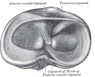
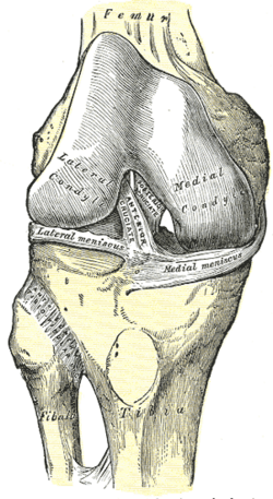
Horns attached to the intercondylar area of the tibia; margins to the capsule (coronary ligaments); the transverse ligament joins them anteriorly. Medial meniscus: C-shaped, wider posteriorly, firmly attached to the MCL — less mobile. Lateral meniscus: nearly circular (O-shaped), not attached to the LCL, more mobile; popliteus attaches to it and pulls it backward during flexion. Meniscofemoral ligaments join the lateral meniscus to the medial femoral condyle alongside the PCL.
- Function
Increase joint congruity, transmit and distribute load (about half of the load across the knee), absorb shock, contribute to stability and lubrication.
- Relations
Blood supply comes from the genicular arteries to the outer third (red zone), which can heal; the inner two-thirds (white zone) is avascular and nourished by synovial fluid, so tears there rarely heal.
Swipe sideways to see the whole diagram →


Clinical correlations
Meniscal tear PANCE
- Mechanism / lesion
Traumatic: twisting on a flexed, weight-bearing knee in young athletes (the medial meniscus is torn more often). Degenerative: minimal trauma (squatting) in middle-aged and older adults, often with OA.
- Classic presentation
Joint-line pain and tenderness, swelling developing gradually over a day (less vascular than an ACL tear), and mechanical symptoms — catching or locking (a displaced bucket-handle tear can block full extension). Positive McMurray, Thessaly and Apley tests.
- Notes
Degenerative tears are treated with physical therapy first (arthroscopy adds little). A locked knee or repairable peripheral tear in a young patient → arthroscopic repair or partial meniscectomy.
Discoid lateral meniscus
- Mechanism / lesion
Congenital disc-shaped lateral meniscus, prone to tearing.
- Classic presentation
"Snapping knee" in a child, sometimes with pain or locking.
High-yield PANCE pearls
- Twisting injury, joint-line tenderness, delayed swelling, locking → meniscal tear.
- Medial meniscus torn more often (attached to the MCL).
- Locked knee (can't fully extend) → bucket-handle tear → urgent ortho/arthroscopy.
- Outer third (red zone) can heal; inner two-thirds can't.
Imaging anatomy
- X-ray
Usually normal; weight-bearing films assess coexisting OA.
- MRI
Test of choice — high signal reaching the articular surface on two slices; the "double PCL" sign of a displaced bucket-handle tear.
- Key views & landmarks
- MRI knee (sagittal and coronal proton-density)
Physical exam
- Surface landmarks
The medial and lateral joint lines are palpable on either side of the patellar ligament with the knee flexed 90°.
- Palpation
Joint-line tenderness is one of the more sensitive findings for meniscal tears.
- Strength / function testing
Check whether the knee fully extends (a mechanical block suggests a displaced tear).
McMurray test
- Technique
Supine; fully flex the knee, then extend it while rotating the tibia (external rotation with valgus for the medial meniscus; internal rotation with varus for the lateral), with fingers on the joint line.
- Positive test
A painful click or clunk at the joint line.
- Significance
Meniscal tear; specific but not very sensitive.
Thessaly test
- Technique
Standing on the affected leg with the knee flexed 20° (holding the examiner's hands), the patient rotates the body and knee internally and externally three times.
- Positive test
Joint-line pain or a sense of locking or catching.
- Significance
Meniscal tear. verify
Apley grind test
- Technique
Prone with the knee flexed 90°; press the heel down (compressing the joint) while rotating the tibia.
- Positive test
Pain that is relieved when the leg is pulled up instead (distraction).
- Significance
Meniscal tear (pain with distraction suggests a ligament problem).
Self-quiz
A 24-year-old man twisted his right knee while playing basketball. Swelling developed the next day. He now reports the knee "catches" and sometimes locks. There is tenderness over the medial joint line. Which structure is most likely injured?
Correct answer: B
Delayed swelling, mechanical symptoms (catching, locking) and joint-line tenderness point to a meniscal tear, most often medial. ACL tears typically cause rapid hemarthrosis within hours.
Show answer
B
Delayed swelling, mechanical symptoms (catching, locking) and joint-line tenderness point to a meniscal tear, most often medial. ACL tears typically cause rapid hemarthrosis within hours.
A 58-year-old woman with knee OA has a degenerative medial meniscal tear on MRI without locking. What is the most appropriate initial treatment?
Correct answer: B
For degenerative tears without a mechanical block, trials show arthroscopic meniscectomy offers little benefit over physical therapy, which is first-line.
Show answer
B
For degenerative tears without a mechanical block, trials show arthroscopic meniscectomy offers little benefit over physical therapy, which is first-line.
Which part of a meniscus is able to heal after a tear?
Correct answer: A
The outer third (red zone) is vascularized by the genicular arteries and can heal. The inner two-thirds is avascular, nourished by synovial fluid, so tears there rarely heal.
Show answer
A
The outer third (red zone) is vascularized by the genicular arteries and can heal. The inner two-thirds is avascular, nourished by synovial fluid, so tears there rarely heal.
A 19-year-old twisted his knee and now cannot fully extend it; the knee is "stuck" in slight flexion. What is the most appropriate management?
Correct answer: C
A locked knee that can't fully extend suggests a displaced bucket-handle meniscal tear, which needs urgent orthopedic referral and arthroscopy (repair or partial meniscectomy). Physical therapy first is for degenerative tears without locking.
Show answer
C
A locked knee that can't fully extend suggests a displaced bucket-handle meniscal tear, which needs urgent orthopedic referral and arthroscopy (repair or partial meniscectomy). Physical therapy first is for degenerative tears without locking.
After a twisting knee injury, which feature favors a meniscal tear over an ACL tear?
Correct answer: D
The menisci are less vascular than the ACL, so swelling from a meniscal tear develops gradually over about a day, with joint-line tenderness and mechanical symptoms (catching or locking). Rapid hemarthrosis within hours suggests an ACL tear.
Show answer
D
The menisci are less vascular than the ACL, so swelling from a meniscal tear develops gradually over about a day, with joint-line tenderness and mechanical symptoms (catching or locking). Rapid hemarthrosis within hours suggests an ACL tear.
Flashcards
See also
- Knee joint
- Collateral ligaments of the knee
- Cruciate ligaments
- Knee ligament and meniscal injuries (comparison table)
- A pop in the knee on landing (clinical case)