Intercostal space
Space between adjacent ribs containing three muscle layers and the intercostal neurovascular bundle, which runs along the lower border of the rib above.
Core anatomy
- Location
Between adjacent ribs, eleven spaces on each side.
- Boundaries
- superior
- Inferior border of the rib above (with its costal groove)
- inferior
- Superior border of the rib below
- superficial
- Skin, subcutaneous tissue, and overlying muscles (pectoral, serratus anterior, back muscles)
- deep
- Endothoracic fascia and parietal pleura
- Contents
- External intercostal muscle: fibers run inferoanteriorly (like hands in front pockets); replaced anteriorly by the external intercostal membrane
- Internal intercostal muscle: fibers run at right angles to the externals; replaced posteriorly by the internal intercostal membrane
- Innermost intercostal muscle (with subcostales and transversus thoracis in the same layer)
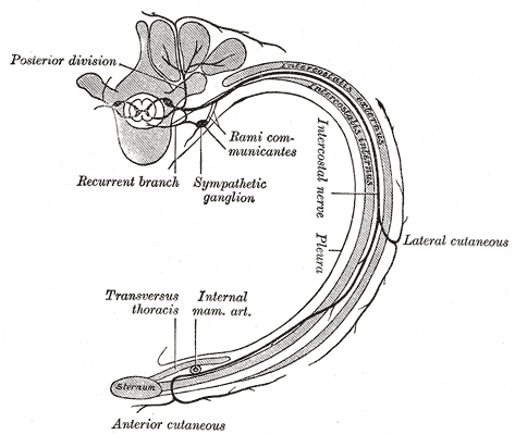
- Intercostal neurovascular bundle: from superior to inferior, Vein, Artery, Nerve (VAN), in the costal groove between the internal and innermost layers
- Collateral branches of the vessels and nerve along the upper border of the rib below
- Communications
Posteriorly with the paravertebral space. The intercostal nerves continue anteriorly into the abdominal wall (T7–T11).
- Notes
Intercostal nerves are the ventral rami of T1–T11 (T12 is the subcostal nerve). They supply the intercostal muscles and a band of skin (dermatome); T4 ≈ nipple, T10 ≈ umbilicus. Posterior intercostal arteries come from the thoracic aorta (spaces 3–11) and the supreme intercostal artery (spaces 1–2). Anterior intercostal arteries come from the internal thoracic artery (spaces 1–6) and the musculophrenic artery. Posterior intercostal veins drain to the azygos vein system.

Clinical correlations
Needle and tube placement (thoracentesis, chest tube) PANCE
- Mechanism / lesion
The main neurovascular bundle lies under the lower border of each rib.
- Classic presentation
Insert needles and tubes just above the upper border of the rib (the lower rib of the space) to avoid the intercostal vein, artery and nerve. Posteriorly, near the angle, the bundle is less protected by the costal groove, so avoid going too medial. Intercostal artery injury can cause significant hemothorax.
Herpes zoster (shingles) PANCE
- Mechanism / lesion
Reactivation of varicella-zoster virus in a dorsal root ganglion, spreading along the intercostal nerve.
- Classic presentation
Burning pain followed by a vesicular rash in a dermatomal band that doesn't cross the midline. Thoracic dermatomes are the most commonly affected.
Intercostal nerve block
- Mechanism / lesion
Local anesthetic injected near the intercostal nerve at the inferior border of the rib, often at the angle.
- Classic presentation
Analgesia for rib fractures or thoracic surgery. Blocking adjacent spaces improves coverage because of overlap.
High-yield PANCE pearls
- Neurovascular bundle order, top to bottom: VAN (vein, artery, nerve), under the rib above.
- Thoracentesis / chest tube: go over the top of the lower rib.
- Dermatomes: T4 nipple, T10 umbilicus.
- Herpes zoster: unilateral dermatomal vesicles; the thoracic region is most common.
Imaging anatomy
- X-ray
Intercostal spaces are counted on the chest X-ray to describe lesion location and line or tube positions.
- CT
Shows the intercostal muscles, vessels (with contrast) and pleura. Useful for chest wall masses and hemothorax from intercostal artery bleeding.
- Ultrasound
Essential for pleural procedures: identify the ribs (acoustic shadows), the pleural line, and fluid; confirm the diaphragm position; color Doppler can show the intercostal artery before needle insertion.
- Key views & landmarks
- Ultrasound in the longitudinal plane across two ribs ("bat sign": ribs as wings, pleural line as body)
Physical exam
- Surface landmarks
Identify the 2nd intercostal space just below the sternal angle and count down. Posteriorly, the inferior angle of the scapula marks about the 7th rib or intercostal space.
- Palpation
Palpate the spaces for tenderness, bulging (effusion, rarely) or retraction.
- Strength / function testing
Map sensory deficits or zoster rashes to dermatomes. Observe intercostal retractions (increased work of breathing).
Self-quiz
During thoracentesis, a needle is inserted just below the inferior border of a rib. Which structure is most at risk?
Correct answer: B
The intercostal vein, artery and nerve run in the costal groove along the inferior border of each rib. Needles should pass just above the superior border of the rib below.
Show answer
B
The intercostal vein, artery and nerve run in the costal groove along the inferior border of each rib. Needles should pass just above the superior border of the rib below.
What is the order of the intercostal neurovascular bundle from superior to inferior?
Correct answer: C
From superior to inferior within the costal groove: Vein, Artery, Nerve (VAN).
Show answer
C
From superior to inferior within the costal groove: Vein, Artery, Nerve (VAN).
A 72-year-old man has burning pain followed by a vesicular band-like rash at the level of his umbilicus on one side. Which dermatome is involved?
Correct answer: C
The umbilicus corresponds to the T10 dermatome. T4 is at the nipple. Herpes zoster follows a single dermatome and doesn't cross the midline.
Show answer
C
The umbilicus corresponds to the T10 dermatome. T4 is at the nipple. Herpes zoster follows a single dermatome and doesn't cross the midline.
The anterior intercostal arteries of spaces 1–6 arise from which artery?
Correct answer: A
The internal thoracic artery gives the anterior intercostal arteries of spaces 1–6; the musculophrenic artery supplies the lower spaces. The posterior intercostal arteries come from the thoracic aorta (spaces 3–11) and the supreme intercostal artery (spaces 1–2).
Show answer
A
The internal thoracic artery gives the anterior intercostal arteries of spaces 1–6; the musculophrenic artery supplies the lower spaces. The posterior intercostal arteries come from the thoracic aorta (spaces 3–11) and the supreme intercostal artery (spaces 1–2).
Which intercostal muscle has fibers running inferoanteriorly, like hands in front pockets?
Correct answer: B
External intercostal fibers run inferoanteriorly ("hands in front pockets"). The internal intercostals run at right angles to them. The neurovascular bundle lies between the internal and innermost layers.
Show answer
B
External intercostal fibers run inferoanteriorly ("hands in front pockets"). The internal intercostals run at right angles to them. The neurovascular bundle lies between the internal and innermost layers.
Flashcards
See also
- Ribs
- Pleural cavity
- Stab wound to the chest (clinical case)